3U81 | pdb_00003u81

Crystal structure of a SAH-bound semi-holo form of rat Catechol-O-methyltransferase


Experimental Data Snapshot

  • Method: X-RAY DIFFRACTION
  • Resolution: 1.13 Å
  • R-Value Free: 
    0.160 (Depositor), 0.160 (DCC) 
  • R-Value Work: 
    0.134 (Depositor), 0.135 (DCC) 
  • R-Value Observed: 
    0.136 (Depositor) 

wwPDB Validation 3D Report Full Report

Validation slider image for 3U81

Ligand Structure Quality Assessment 


This is version 1.3 of the entry. See complete history

Literature

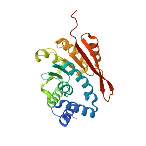
Catechol-O-methyltransferase in complex with substituted 3'-deoxyribose bisubstrate inhibitors.

Ellermann, M.Lerner, C.Burgy, G.Ehler, A.Bissantz, C.Jakob-Roetne, R.Paulini, R.Allemann, O.Tissot, H.Grunstein, D.Stihle, M.Diederich, F.Rudolph, M.G.

(2012) Acta Crystallogr D Biol Crystallogr 68: 253-260

  • DOI: https://doi.org/10.1107/S0907444912001138
  • Primary Citation Related Structures: 
    3NWB, 3NWE, 3R6T, 3S68, 3U81

  • PubMed Abstract: 

    The biological activity of catechol neurotransmitters such as dopamine in the synapse is modulated by transporters and enzymes. Catechol-O-methyltransferase (COMT; EC 2.1.1.6) inactivates neurotransmitters by catalyzing the transfer of a methyl group from S-adenosylmethionine to catechols in the presence of Mg²⁺. This pathway also inactivates L-DOPA, the standard therapeutic for Parkinson's disease. Depletion of catechol neurotransmitters in the prefrontal cortex has been linked to schizophrenia. The inhibition of COMT therefore promises improvements in the treatment of these diseases. The concept of bisubstrate inhibitors for COMT has been described previously. Here, ribose-modified bisubstrate inhibitors were studied. Three high-resolution crystal structures of COMT in complex with novel ribose-modified bisubstrate inhibitors confirmed the predicted binding mode but displayed subtle alterations at the ribose-binding site. The high affinity of the inhibitors can be convincingly rationalized from the structures, which document the possibility of removing and/or replacing the ribose 3'-hydroxyl group and provide a framework for further inhibitor design.


  • Organizational Affiliation
    • Laboratorium für Organische Chemie, ETH Zürich, Hönggerberg, HCI, CH-8093 Zürich, Switzerland.

Macromolecule Content 

  • Total Structure Weight: 25.12 kDa 
  • Atom Count: 2,145 
  • Modeled Residue Count: 218 
  • Deposited Residue Count: 221 
  • Unique protein chains: 1

Macromolecules

Find similar proteins by:|  3D Structure
Entity ID: 1
MoleculeChains  Sequence LengthOrganismDetailsImage
Catechol O-methyltransferase221Rattus norvegicusMutation(s): 2 
Gene Names: Comt
EC: 2.1.1.6
UniProt
Find proteins for P22734 (Rattus norvegicus)
Explore P22734 
Go to UniProtKB:  P22734
Entity Groups
Sequence Clusters30% Identity50% Identity70% Identity90% Identity95% Identity100% Identity
UniProt GroupP22734
Sequence Annotations
Expand
Reference Sequence

Small Molecules

Experimental Data & Validation

Experimental Data

  • Method: X-RAY DIFFRACTION
  • Resolution: 1.13 Å
  • R-Value Free:  0.160 (Depositor), 0.160 (DCC) 
  • R-Value Work:  0.134 (Depositor), 0.135 (DCC) 
  • R-Value Observed: 0.136 (Depositor) 
Space Group: P 21 21 21
Unit Cell:
Length ( Å )Angle ( ˚ )
a = 33.384α = 90
b = 61.333β = 90
c = 104.686γ = 90
Software Package:
Software NamePurpose
PHASERphasing
PHENIXrefinement
XDSdata reduction
SADABSdata scaling

Structure Validation

View Full Validation Report


Ligand Structure Quality Assessment 


Entry History 

Deposition Data

Revision History  (Full details and data files)

  • Version 1.0: 2012-02-01
    Type: Initial release
  • Version 1.1: 2012-04-11
    Changes: Database references
  • Version 1.2: 2017-11-08
    Changes: Refinement description
  • Version 1.3: 2024-02-28
    Changes: Data collection, Database references, Derived calculations