3U7B | pdb_00003u7b

A new crystal structure of a Fusarium oxysporum GH10 xylanase reveals the presence of an extended loop on top of the catalytic cleft


Experimental Data Snapshot

  • Method: X-RAY DIFFRACTION
  • Resolution: 1.94 Å
  • R-Value Free: 
    0.242 (Depositor), 0.240 (DCC) 
  • R-Value Work: 
    0.213 (Depositor), 0.212 (DCC) 
  • R-Value Observed: 
    0.214 (Depositor) 

Starting Model: experimental
View more details

wwPDB Validation 3D Report Full Report

Validation slider image for 3U7B

This is version 2.2 of the entry. See complete history

Literature

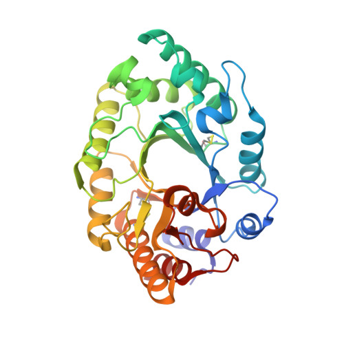
The structure of a GH10 xylanase from Fusarium oxysporum reveals the presence of an extended loop on top of the catalytic cleft.

Dimarogona, M.Topakas, E.Christakopoulos, P.Chrysina, E.D.

(2012) Acta Crystallogr D Biol Crystallogr 68: 735-742

  • DOI: https://doi.org/10.1107/S0907444912007044
  • Primary Citation Related Structures: 
    3U7B

  • PubMed Abstract: 

    Xylanase enzymes have been the focus of considerable research in recent decades owing to their extensive use in a variety of biotechnological applications. Previous structural studies of a number of GH10 xylanases revealed that all GH10 family members have the (β/α)(8)-barrel fold and their catalytic site is conserved. The structure of a new GH10 xylanase from Fusarium oxysporum (FoXyn10a) was determined at 1.94 Å resolution from crystals belonging to the tetragonal space group P4(1)2(1)2 with five molecules per asymmetric unit. Comparison of the structure of FoXyn10a with previously determined structures of GH10 family members indicated that most of the differences were located in the loop regions between the ordered secondary-structure elements of the barrel, as expected. However, alignment of FoXyn10a with sequence and structural homologues denoted an atypically long loop connecting strand β6b and helix α6 that was only present in one other GH10 xylanase, the structure of which is not known. This structural feature may be of functional importance, with potential implications in the catalytic efficiency of the enzyme.


  • Organizational Affiliation
    • Institute of Organic and Pharmaceutical Chemistry, National Hellenic Research Foundation, 48 Vassileos Constantinou Avenue, 11635 Athens, Greece.

Macromolecule Content 

  • Total Structure Weight: 186.38 kDa 
  • Atom Count: 14,970 
  • Modeled Residue Count: 1,632 
  • Deposited Residue Count: 1,635 
  • Unique protein chains: 1

Macromolecules

Find similar proteins by:|  3D Structure
Entity ID: 1
MoleculeChains  Sequence LengthOrganismDetailsImage
ENDO-1,4-BETA-XYLANASE
A, B, C, D, E
327Fusarium oxysporumMutation(s): 0 
EC: 3.2.1.8
UniProt
Find proteins for B3A0S5 (Fusarium oxysporum f. sp. lycopersici (strain 4287 / CBS 123668 / FGSC 9935 / NRRL 34936))
Explore B3A0S5 
Go to UniProtKB:  B3A0S5
Entity Groups
Sequence Clusters30% Identity50% Identity70% Identity90% Identity95% Identity100% Identity
UniProt GroupB3A0S5
Glycosylation
Glycosylation Sites: 1
Sequence Annotations
Expand
Reference Sequence

Oligosaccharides

Help  
Entity ID: 2
MoleculeChains Length2D Diagram GlycosylationD Interactions
beta-D-mannopyranose-(1-4)-2-acetamido-2-deoxy-beta-D-glucopyranose-(1-4)-2-acetamido-2-deoxy-beta-D-glucopyranose
F, H
3N-Glycosylation
Glycosylation Resources
GlyTouCan: G15407YE
GlyCosmos: G15407YE
GlyGen: G15407YE
Entity ID: 3
MoleculeChains Length2D Diagram GlycosylationD Interactions
alpha-D-mannopyranose-(1-2)-alpha-D-mannopyranose-(1-3)-beta-D-mannopyranose-(1-4)-2-acetamido-2-deoxy-beta-D-glucopyranose-(1-4)-2-acetamido-2-deoxy-beta-D-glucopyranose
G
5N-Glycosylation
Glycosylation Resources
GlyTouCan: G42227JK
GlyCosmos: G42227JK
GlyGen: G42227JK
Entity ID: 4
MoleculeChains Length2D Diagram GlycosylationD Interactions
alpha-D-mannopyranose-(1-6)-beta-D-mannopyranose-(1-4)-2-acetamido-2-deoxy-beta-D-glucopyranose-(1-4)-2-acetamido-2-deoxy-beta-D-glucopyranose
I, J
4N-Glycosylation
Glycosylation Resources
GlyTouCan: G22573RC
GlyCosmos: G22573RC
GlyGen: G22573RC

Small Molecules

Ligands 1 Unique
IDChains Name / Formula / InChI Key2D Diagram3D Interactions
EDO

Query on EDO


Download:Ideal Coordinates CCD File
AA [auth C]
BA [auth C]
CA [auth C]
DA [auth C]
EA [auth D]
AA [auth C],
BA [auth C],
CA [auth C],
DA [auth C],
EA [auth D],
FA [auth D],
GA [auth D],
HA [auth E],
IA [auth E],
JA [auth E],
K [auth A],
L [auth A],
M [auth A],
N [auth A],
O [auth A],
P [auth A],
Q [auth A],
R [auth B],
S [auth B],
T [auth B],
U [auth B],
V [auth B],
W [auth B],
X [auth B],
Y [auth C],
Z [auth C]
1,2-ETHANEDIOL
C2 H6 O2
LYCAIKOWRPUZTN-UHFFFAOYSA-N

Experimental Data & Validation

Experimental Data

  • Method: X-RAY DIFFRACTION
  • Resolution: 1.94 Å
  • R-Value Free:  0.242 (Depositor), 0.240 (DCC) 
  • R-Value Work:  0.213 (Depositor), 0.212 (DCC) 
  • R-Value Observed: 0.214 (Depositor) 
Space Group: P 41 21 2
Unit Cell:
Length ( Å )Angle ( ˚ )
a = 124.22α = 90
b = 124.22β = 90
c = 284.04γ = 90
Software Package:
Software NamePurpose
DNAdata collection
BALBESphasing
REFMACrefinement
MOSFLMdata reduction
SCALAdata scaling

Structure Validation

View Full Validation Report


Entry History 

Deposition Data

Revision History  (Full details and data files)

  • Version 1.0: 2012-07-18
    Type: Initial release
  • Version 2.0: 2020-07-29
    Type: Remediation
    Reason: Carbohydrate remediation
    Changes: Advisory, Atomic model, Data collection, Derived calculations, Structure summary
  • Version 2.1: 2023-09-13
    Changes: Data collection, Database references, Refinement description, Structure summary
  • Version 2.2: 2024-11-06
    Changes: Structure summary