3U5M | pdb_00003u5m

Crystal structure of TRIM33 PHD-Bromo in the free state


Experimental Data Snapshot

  • Method: X-RAY DIFFRACTION
  • Resolution: 3.08 Å
  • R-Value Free: 
    0.293 (Depositor), 0.286 (DCC) 
  • R-Value Work: 
    0.207 (Depositor), 0.193 (DCC) 
  • R-Value Observed: 
    0.211 (Depositor) 

Starting Model: experimental
View more details

wwPDB Validation 3D Report Full Report

Validation slider image for 3U5M

This is version 1.1 of the entry. See complete history

Literature

A poised chromatin platform for TGF-beta access to master regulators

Xi, Q.Wang, Z.Zaromytidou, A.I.Zhang, X.H.Chow-Tsang, L.F.Liu, J.X.Kim, H.Barlas, A.Manova-Todorova, K.Kaartinen, V.Studer, L.Mark, W.Patel, D.J.Massague, J.

(2011) Cell 147: 1511-1524

  • DOI: https://doi.org/10.1016/j.cell.2011.11.032
  • Primary Citation Related Structures: 
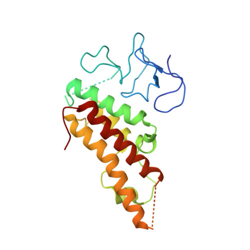
    3U5M, 3U5N, 3U5O, 3U5P

  • PubMed Abstract: 

    Specific chromatin marks keep master regulators of differentiation silent yet poised for activation by extracellular signals. We report that nodal TGF-β signals use the poised histone mark H3K9me3 to trigger differentiation of mammalian embryonic stem cells. Nodal receptors induce the formation of companion Smad4-Smad2/3 and TRIM33-Smad2/3 complexes. The PHD-Bromo cassette of TRIM33 facilitates binding of TRIM33-Smad2/3 to H3K9me3 and H3K18ac on the promoters of mesendoderm regulators Gsc and Mixl1. The crystal structure of this cassette, bound to histone H3 peptides, illustrates that PHD recognizes K9me3, and Bromo binds an adjacent K18ac. The interaction between TRIM33-Smad2/3 and H3K9me3 displaces the chromatin-compacting factor HP1γ, making nodal response elements accessible to Smad4-Smad2/3 for Pol II recruitment. In turn, Smad4 increases K18 acetylation to augment TRIM33-Smad2/3 binding. Thus, nodal effectors use the H3K9me3 mark as a platform to switch master regulators of stem cell differentiation from the poised to the active state.


  • Organizational Affiliation
    • Cancer Biology and Genetics Program, Memorial Sloan-Kettering Cancer Center, New York, NY 10065, USA.

Macromolecule Content 

  • Total Structure Weight: 288.01 kDa 
  • Atom Count: 17,117 
  • Modeled Residue Count: 2,114 
  • Deposited Residue Count: 2,484 
  • Unique protein chains: 1

Macromolecules

Find similar proteins by:|  3D Structure
Entity ID: 1
MoleculeChains  Sequence LengthOrganismDetailsImage
E3 ubiquitin-protein ligase TRIM33
A, B, C, D, E
A, B, C, D, E, F, G, H, I, J, K, L
207Homo sapiensMutation(s): 0 
Gene Names: TRIM33KIAA1113RFG7TIF1G
EC: 6.3.2 (PDB Primary Data), 2.3.2.27 (UniProt)
UniProt & NIH Common Fund Data Resources
Find proteins for Q9UPN9 (Homo sapiens)
Explore Q9UPN9 
Go to UniProtKB:  Q9UPN9
PHAROS:  Q9UPN9
GTEx:  ENSG00000197323 
Entity Groups
Sequence Clusters30% Identity50% Identity70% Identity90% Identity95% Identity100% Identity
UniProt GroupQ9UPN9
Sequence Annotations
Expand
Reference Sequence

Small Molecules

Ligands 2 Unique
IDChains Name / Formula / InChI Key2D Diagram3D Interactions
ZN

Query on ZN


Download:Ideal Coordinates CCD File
BA [auth E]
CA [auth E]
EA [auth F]
FA [auth F]
IA [auth G]
BA [auth E],
CA [auth E],
EA [auth F],
FA [auth F],
IA [auth G],
JA [auth G],
KA [auth H],
LA [auth H],
M [auth A],
MA [auth I],
N [auth A],
NA [auth I],
Q [auth B],
QA [auth J],
R [auth B],
RA [auth J],
SA [auth K],
TA [auth K],
U [auth C],
UA [auth L],
V [auth C],
VA [auth L],
Y [auth D],
Z [auth D]
ZINC ION
Zn
PTFCDOFLOPIGGS-UHFFFAOYSA-N
CA

Query on CA


Download:Ideal Coordinates CCD File
AA [auth E]
DA [auth E]
GA [auth F]
HA [auth F]
O [auth A]
AA [auth E],
DA [auth E],
GA [auth F],
HA [auth F],
O [auth A],
OA [auth I],
P [auth A],
PA [auth I],
S [auth B],
T [auth B],
W [auth C],
X [auth C]
CALCIUM ION
Ca
BHPQYMZQTOCNFJ-UHFFFAOYSA-N

Experimental Data & Validation

Experimental Data

  • Method: X-RAY DIFFRACTION
  • Resolution: 3.08 Å
  • R-Value Free:  0.293 (Depositor), 0.286 (DCC) 
  • R-Value Work:  0.207 (Depositor), 0.193 (DCC) 
  • R-Value Observed: 0.211 (Depositor) 
Space Group: P 1
Unit Cell:
Length ( Å )Angle ( ˚ )
a = 79.527α = 89.9
b = 79.781β = 89.96
c = 134.16γ = 59.97
Software Package:
Software NamePurpose
MAR345dtbdata collection
PHASERphasing
PHENIXrefinement
HKL-2000data reduction
HKL-2000data scaling

Structure Validation

View Full Validation Report


Entry History 

Deposition Data

Revision History  (Full details and data files)

  • Version 1.0: 2012-01-18
    Type: Initial release
  • Version 1.1: 2023-09-13
    Changes: Data collection, Database references, Derived calculations, Refinement description