3U54 | pdb_00003u54

Crystal structure (Type-1) of SAICAR synthetase from Pyrococcus horikoshii OT3


Experimental Data Snapshot

  • Method: X-RAY DIFFRACTION
  • Resolution: 2.35 Å
  • R-Value Free: 
    0.286 (Depositor), 0.283 (DCC) 
  • R-Value Work: 
    0.235 (Depositor), 0.232 (DCC) 
  • R-Value Observed: 
    0.237 (Depositor) 

Starting Model: experimental
View more details

wwPDB Validation 3D Report Full Report

Validation slider image for 3U54

This is version 1.4 of the entry. See complete history

Literature

Structure of SAICAR synthetase from Pyrococcus horikoshii OT3: insights into thermal stability

Manjunath, K.Kanaujia, S.P.Kanagaraj, S.Jeyakanthan, J.Sekar, K.

(2013) Int J Biol Macromol 53: 7-19

  • DOI: https://doi.org/10.1016/j.ijbiomac.2012.10.028
  • Primary Citation Related Structures: 
    3U54, 3U55

  • PubMed Abstract: 

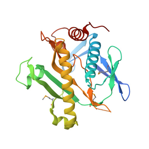
    The first native crystal structure of Phosphoribosylaminoimidazole-succinocarboxamide synthetase (SAICAR synthetase) from a hyperthermophilic organism Pyrococcus horikoshii OT3 was determined in two space groups H3 (Type-1: Resolution 2.35Å) and in C222(1) (Type-2: Resolution 1.9Å). Both are dimeric but Type-1 structure exhibited hexameric arrangement due to the presence of cadmium ions. A comparison has been made on the sequence and structures of all SAICAR synthetases to better understand the differences between mesophilic, thermophilic and hyperthermophilic SAICAR synthetases. These SAICAR synthetases are reasonably similar in sequence and three-dimensional structure; however, differences were visible only in the subtler details of percentage composition of the sequences, salt bridge interactions and non-polar contact areas.


  • Organizational Affiliation
    • Supercomputer Education and Research Centre, Indian Institute of Science, Bangalore 560 012, India.

Macromolecule Content 

  • Total Structure Weight: 57.31 kDa 
  • Atom Count: 3,740 
  • Modeled Residue Count: 450 
  • Deposited Residue Count: 476 
  • Unique protein chains: 1

Macromolecules

Find similar proteins by:|  3D Structure
Entity ID: 1
MoleculeChains  Sequence LengthOrganismDetailsImage
Phosphoribosylaminoimidazole-succinocarboxamide synthase
A, B
238Pyrococcus horikoshii OT3Mutation(s): 0 
Gene Names: PH0239purC
EC: 6.3.2.6
UniProt
Find proteins for O57978 (Pyrococcus horikoshii (strain ATCC 700860 / DSM 12428 / JCM 9974 / NBRC 100139 / OT-3))
Explore O57978 
Go to UniProtKB:  O57978
Entity Groups
Sequence Clusters30% Identity50% Identity70% Identity90% Identity95% Identity100% Identity
UniProt GroupO57978
Sequence Annotations
Expand
Reference Sequence

Small Molecules

Ligands 4 Unique
IDChains Name / Formula / InChI Key2D Diagram3D Interactions
CD

Query on CD


Download:Ideal Coordinates CCD File
E [auth A]
F [auth A]
G [auth A]
H [auth A]
I [auth A]
E [auth A],
F [auth A],
G [auth A],
H [auth A],
I [auth A],
J [auth A],
L [auth B],
M [auth B],
N [auth B],
O [auth B],
P [auth B],
Q [auth B]
CADMIUM ION
Cd
WLZRMCYVCSSEQC-UHFFFAOYSA-N
SO4

Query on SO4


Download:Ideal Coordinates CCD File
D [auth A],
K [auth A],
R [auth B],
S [auth B]
SULFATE ION
O4 S
QAOWNCQODCNURD-UHFFFAOYSA-L
BU1

Query on BU1


Download:Ideal Coordinates CCD File
C [auth A]1,4-BUTANEDIOL
C4 H10 O2
WERYXYBDKMZEQL-UHFFFAOYSA-N
ACT

Query on ACT


Download:Ideal Coordinates CCD File
T [auth B]ACETATE ION
C2 H3 O2
QTBSBXVTEAMEQO-UHFFFAOYSA-M
Modified Residues  1 Unique
IDChains TypeFormula2D DiagramParent
MSE
Query on MSE
A, B
L-PEPTIDE LINKINGC5 H11 N O2 SeMET

Experimental Data & Validation

Experimental Data

  • Method: X-RAY DIFFRACTION
  • Resolution: 2.35 Å
  • R-Value Free:  0.286 (Depositor), 0.283 (DCC) 
  • R-Value Work:  0.235 (Depositor), 0.232 (DCC) 
  • R-Value Observed: 0.237 (Depositor) 
Space Group: H 3
Unit Cell:
Length ( Å )Angle ( ˚ )
a = 95.42α = 90
b = 95.42β = 90
c = 148.631γ = 120
Software Package:
Software NamePurpose
MAR345dtbdata collection
PHASERphasing
REFMACrefinement
MOSFLMdata reduction
SCALAdata scaling

Structure Validation

View Full Validation Report


Entry History 

Deposition Data

Revision History  (Full details and data files)

  • Version 1.0: 2012-10-17
    Type: Initial release
  • Version 1.1: 2013-12-18
    Changes: Database references
  • Version 1.2: 2023-11-01
    Changes: Data collection, Database references, Derived calculations, Refinement description
  • Version 1.3: 2023-12-06
    Changes: Data collection
  • Version 1.4: 2024-11-13
    Changes: Structure summary