3U48 | pdb_00003u48

From soil to structure: a novel dimeric family 3-beta-glucosidase isolated from compost using metagenomic analysis


Experimental Data Snapshot

  • Method: X-RAY DIFFRACTION
  • Resolution: 2.20 Å
  • R-Value Free: 
    0.219 (Depositor), 0.206 (DCC) 
  • R-Value Work: 
    0.169 (Depositor), 0.161 (DCC) 
  • R-Value Observed: 
    0.169 (Depositor) 

wwPDB Validation 3D Report Full Report

Validation slider image for 3U48

Ligand Structure Quality Assessment 


This is version 1.4 of the entry. See complete history

Literature

From soil to structure, a novel dimeric beta-glucosidase belonging to glycoside hydrolase family 3 isolated from compost using metagenomic analysis.

McAndrew, R.P.Park, J.I.Heins, R.A.Reindl, W.Friedland, G.D.D'haeseleer, P.Northen, T.Sale, K.L.Simmons, B.A.Adams, P.D.

(2013) J Biological Chem 288: 14985-14992

  • DOI: https://doi.org/10.1074/jbc.M113.458356
  • Primary Citation Related Structures: 
    3U48, 3U4A

  • PubMed Abstract: 

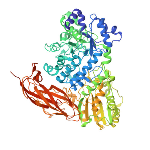
    A recent metagenomic analysis sequenced a switchgrass-adapted compost community to identify enzymes from microorganisms that were specifically adapted to switchgrass under thermophilic conditions. These enzymes are being examined as part of the pretreatment process for the production of "second-generation" biofuels. Among the enzymes discovered was JMB19063, a novel three-domain β-glucosidase that belongs to the GH3 (glycoside hydrolase 3) family. Here, we report the structure of JMB19063 in complex with glucose and the catalytic variant D261N crystallized in the presence of cellopentaose. JMB19063 is first structure of a dimeric member of the GH3 family, and we demonstrate that dimerization is required for catalytic activity. Arg-587 and Phe-598 from the C-terminal domain of the opposing monomer are shown to interact with bound ligands in the D261N structure. Enzyme assays confirmed that these residues are absolutely essential for full catalytic activity.


  • Organizational Affiliation
    • Joint BioEnergy Institute, Emeryville, California 94608, USA.

Macromolecule Content 

  • Total Structure Weight: 171.41 kDa 
  • Atom Count: 12,332 
  • Modeled Residue Count: 1,481 
  • Deposited Residue Count: 1,550 
  • Unique protein chains: 1

Macromolecules

Find similar proteins by:|  3D Structure
Entity ID: 1
MoleculeChains  Sequence LengthOrganismDetailsImage
JMB19063
A, B
775compost metagenomeMutation(s): 0 

Experimental Data & Validation

Experimental Data

  • Method: X-RAY DIFFRACTION
  • Resolution: 2.20 Å
  • R-Value Free:  0.219 (Depositor), 0.206 (DCC) 
  • R-Value Work:  0.169 (Depositor), 0.161 (DCC) 
  • R-Value Observed: 0.169 (Depositor) 
Space Group: P 43 21 2
Unit Cell:
Length ( Å )Angle ( ˚ )
a = 121.488α = 90
b = 121.488β = 90
c = 242.859γ = 90
Software Package:
Software NamePurpose
BOSdata collection
PHENIXmodel building
PHENIXrefinement
HKL-2000data reduction
HKL-2000data scaling
PHENIXphasing

Structure Validation

View Full Validation Report


Ligand Structure Quality Assessment 


Entry History 

Deposition Data

Revision History  (Full details and data files)

  • Version 1.0: 2013-04-10
    Type: Initial release
  • Version 1.1: 2014-04-02
    Changes: Source and taxonomy
  • Version 1.2: 2020-07-29
    Type: Remediation
    Reason: Carbohydrate remediation
    Changes: Data collection, Derived calculations, Structure summary
  • Version 1.3: 2024-02-28
    Changes: Data collection, Database references, Structure summary
  • Version 1.4: 2024-11-20
    Changes: Database references, Structure summary