
 Download Mendeley
A novel compound from a molecular fragment library screen inhibits glycosyltransferases by displacing the metal ion and interfering with substrate binding
Jorgensen, R., Grimm, L.L., Sindhuwinata, N., Peters, T., Palcic, M.M.To be published.
Experimental Data Snapshot
Starting Model: experimental
View more details
Entity ID: 1 | |||||
|---|---|---|---|---|---|
| Molecule | Chains | Sequence Length | Organism | Details | Image |
| Histo-blood group ABO system transferase | 298 | Homo sapiens | Mutation(s): 2 Gene Names: ABO EC: 2.4.1.40 (PDB Primary Data), 2.4.1.37 (PDB Primary Data) | 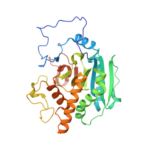 | |
UniProt & NIH Common Fund Data Resources | |||||
PHAROS: P16442 | |||||
Entity Groups | |||||
| Sequence Clusters | 30% Identity50% Identity70% Identity90% Identity95% Identity100% Identity | ||||
| UniProt Group | P16442 | ||||
Sequence AnnotationsExpand | |||||
Reference Sequence | |||||
| Ligands 5 Unique | |||||
|---|---|---|---|---|---|
| ID | Chains | Name / Formula / InChI Key | 2D Diagram | 3D Interactions | |
| UDP Download:Ideal Coordinates CCD File | D [auth A], I [auth B] | URIDINE-5'-DIPHOSPHATE C9 H14 N2 O12 P2 XCCTYIAWTASOJW-XVFCMESISA-N | 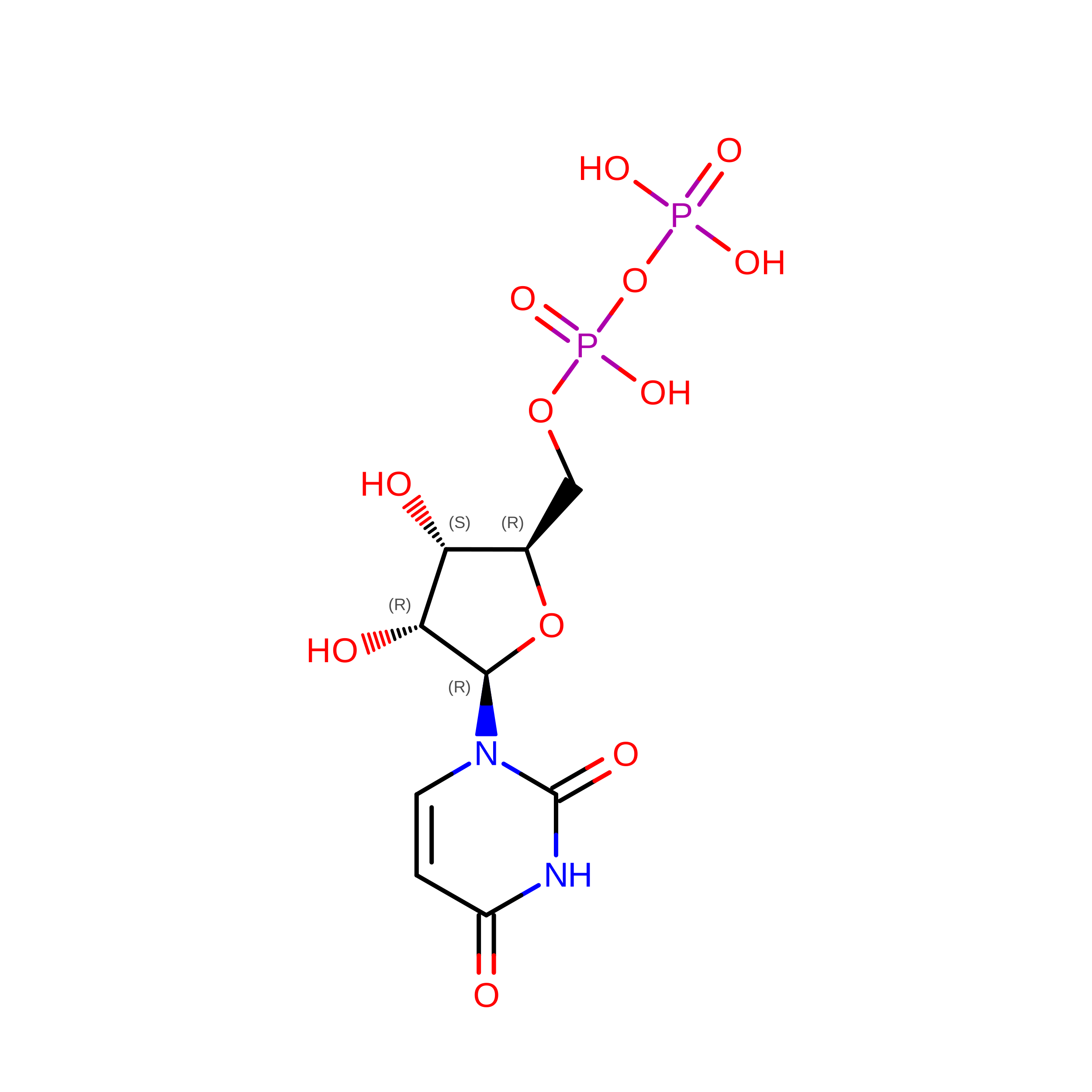 | ||
| GTI Download:Ideal Coordinates CCD File | C [auth A], K [auth B] | 1-(3-phenyl-1,2,4-thiadiazol-5-yl)piperazine C12 H14 N4 S UMFMHSLRNJBGKO-UHFFFAOYSA-N | 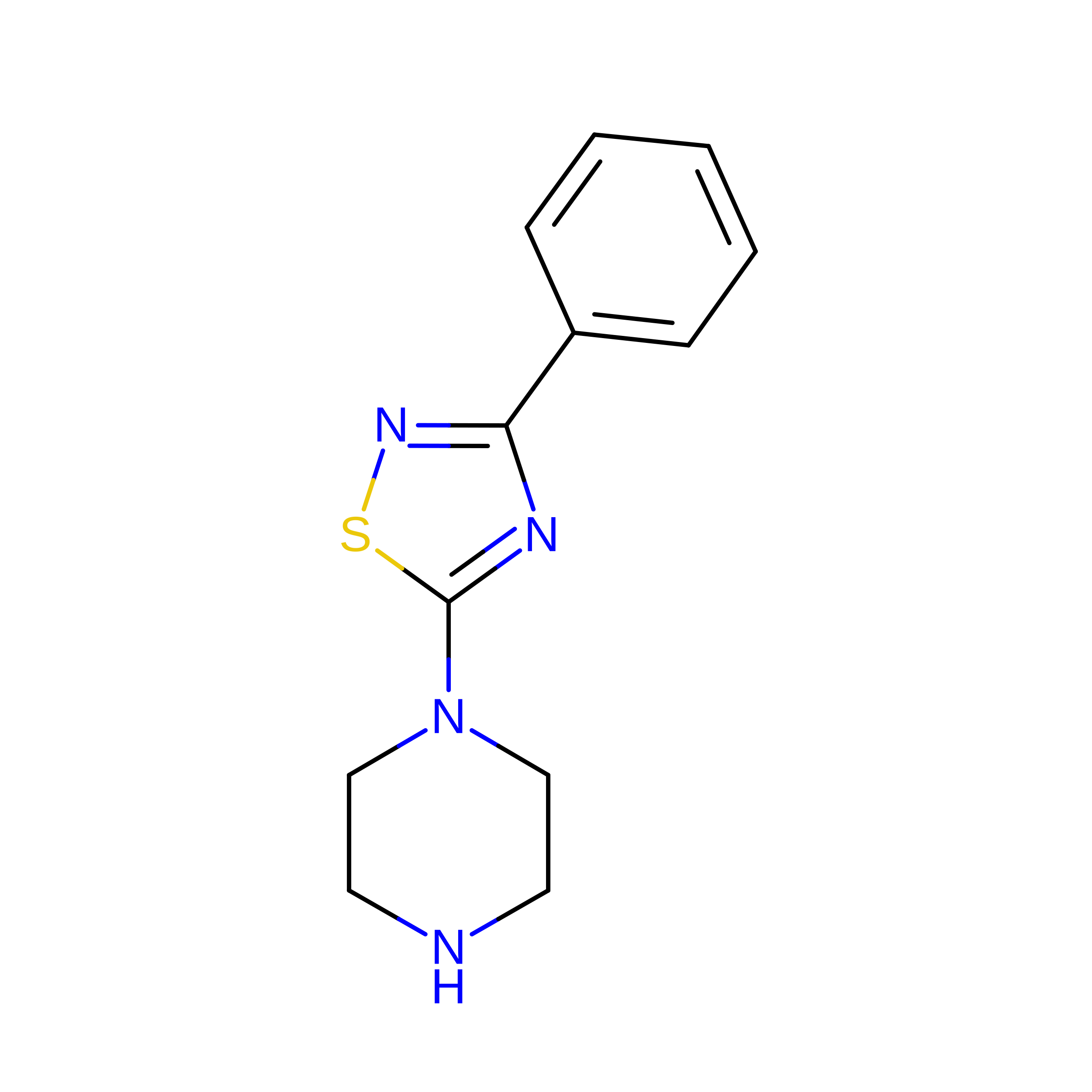 | ||
| SO4 Download:Ideal Coordinates CCD File | F [auth A], H [auth A] | SULFATE ION O4 S QAOWNCQODCNURD-UHFFFAOYSA-L |  | ||
| GOL Download:Ideal Coordinates CCD File | E [auth A], L [auth B], M [auth B] | GLYCEROL C3 H8 O3 PEDCQBHIVMGVHV-UHFFFAOYSA-N |  | ||
| MN Download:Ideal Coordinates CCD File | G [auth A], J [auth B] | MANGANESE (II) ION Mn WAEMQWOKJMHJLA-UHFFFAOYSA-N |  | ||
| Length ( Å ) | Angle ( ˚ ) |
|---|---|
| a = 77.63 | α = 90 |
| b = 152.74 | β = 90 |
| c = 52.99 | γ = 90 |
| Software Name | Purpose |
|---|---|
| XSCALE | data scaling |
| PHASER | phasing |
| PHENIX | refinement |
| PDB_EXTRACT | data extraction |
| MxCuBE | data collection |
