3U0F | pdb_00003u0f

The structure of Beta-ketoacyl synthase from Brucella melitensis bound to the fragment 7-hydroxycoumarin


Experimental Data Snapshot

  • Method: X-RAY DIFFRACTION
  • Resolution: 1.25 Å
  • R-Value Free: 
    0.134 (Depositor), 0.142 (DCC) 
  • R-Value Work: 
    0.122 (Depositor), 0.129 (DCC) 
  • R-Value Observed: 
    0.123 (Depositor) 

Starting Model: experimental
View more details

wwPDB Validation 3D Report Full Report

Validation slider image for 3U0F

Ligand Structure Quality Assessment 


This is version 1.4 of the entry. See complete history

Literature

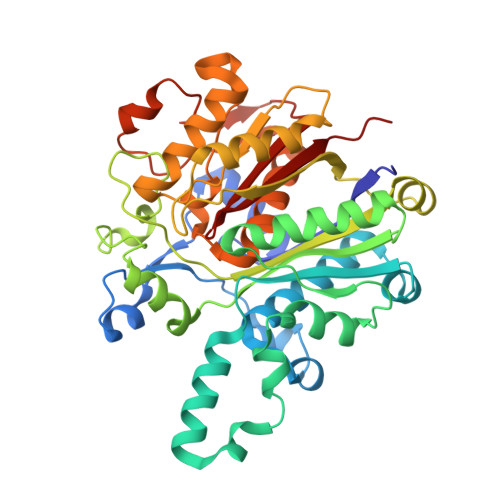
Structural characterization of beta-ketoacyl ACP synthase I bound to platencin and fragment screening molecules at two substrate binding sites.

Patterson, E.I.Nanson, J.D.Abendroth, J.Bryan, C.Sankaran, B.Myler, P.J.Forwood, J.K.

(2020) Proteins 88: 47-56

  • DOI: https://doi.org/10.1002/prot.25765
  • Primary Citation Related Structures: 
    3LRF, 3MQD, 3U0E, 3U0F, 4JV3

  • PubMed Abstract: 

    The bacterial fatty acid pathway is essential for membrane synthesis and a range of other metabolic and cellular functions. The β-ketoacyl-ACP synthases carry out the initial elongation reaction of this pathway, utilizing acetyl-CoA as a primer to elongate malonyl-ACP by two carbons, and subsequent elongation of the fatty acyl-ACP substrate by two carbons. Here we describe the structures of the β-ketoacyl-ACP synthase I from Brucella melitensis in complex with platencin, 7-hydroxycoumarin, and (5-thiophen-2-ylisoxazol-3-yl)methanol. The enzyme is a dimer and based on structural and sequence conservation, harbors the same active site configuration as other β-ketoacyl-ACP synthases. The platencin binding site overlaps with the fatty acyl compound supplied by ACP, while 7-hydroxyl-coumarin and (5-thiophen-2-ylisoxazol-3-yl)methanol bind at the secondary fatty acyl binding site. These high-resolution structures, ranging between 1.25 and 1.70 å resolution, provide a basis for in silico inhibitor screening and optimization, and can aid in rational drug design by revealing the high-resolution binding interfaces of molecules at the malonyl-ACP and acyl-ACP active sites.


  • Organizational Affiliation
    • Department of Pathology, University of Texas Medical Branch, Galveston, Texas.

Macromolecule Content 

  • Total Structure Weight: 43.97 kDa 
  • Atom Count: 3,681 
  • Modeled Residue Count: 407 
  • Deposited Residue Count: 411 
  • Unique protein chains: 1

Macromolecules

Find similar proteins by:|  3D Structure
Entity ID: 1
MoleculeChains  Sequence LengthOrganismDetailsImage
Beta-ketoacyl synthase411Brucella abortus 2308Mutation(s): 0 
Gene Names: BAB1_2173fabB
EC: 2.3.1.41
UniProt
Find proteins for Q2YQQ9 (Brucella abortus (strain 2308))
Explore Q2YQQ9 
Go to UniProtKB:  Q2YQQ9
Entity Groups
Sequence Clusters30% Identity50% Identity70% Identity90% Identity95% Identity100% Identity
UniProt GroupQ2YQQ9
Sequence Annotations
Expand
Reference Sequence

Small Molecules

Ligands 4 Unique
IDChains Name / Formula / InChI Key2D Diagram3D Interactions
07L

Query on 07L


Download:Ideal Coordinates CCD File
E [auth A]7-hydroxy-2H-chromen-2-one
C9 H6 O3
ORHBXUUXSCNDEV-UHFFFAOYSA-N
CL

Query on CL


Download:Ideal Coordinates CCD File
B [auth A],
C [auth A]
CHLORIDE ION
Cl
VEXZGXHMUGYJMC-UHFFFAOYSA-M
MOH

Query on MOH


Download:Ideal Coordinates CCD File
F [auth A],
G [auth A]
METHANOL
C H4 O
OKKJLVBELUTLKV-UHFFFAOYSA-N
NA

Query on NA


Download:Ideal Coordinates CCD File
D [auth A]SODIUM ION
Na
FKNQFGJONOIPTF-UHFFFAOYSA-N

Experimental Data & Validation

Experimental Data

  • Method: X-RAY DIFFRACTION
  • Resolution: 1.25 Å
  • R-Value Free:  0.134 (Depositor), 0.142 (DCC) 
  • R-Value Work:  0.122 (Depositor), 0.129 (DCC) 
  • R-Value Observed: 0.123 (Depositor) 
Space Group: C 1 2 1
Unit Cell:
Length ( Å )Angle ( ˚ )
a = 78.56α = 90
b = 83.25β = 121.2
c = 73.81γ = 90
Software Package:
Software NamePurpose
XSCALEdata scaling
REFMACrefinement
PDB_EXTRACTdata extraction
XDSdata reduction
REFMACphasing

Structure Validation

View Full Validation Report


Ligand Structure Quality Assessment 


Entry History 

Deposition Data

Revision History  (Full details and data files)

  • Version 1.0: 2011-10-12
    Type: Initial release
  • Version 1.1: 2019-08-21
    Changes: Data collection, Database references, Derived calculations
  • Version 1.2: 2023-07-26
    Changes: Database references, Derived calculations
  • Version 1.3: 2023-09-13
    Changes: Data collection, Refinement description
  • Version 1.4: 2025-10-22
    Changes: Structure summary