3TZW | pdb_00003tzw

Crystal structure of a fragment containing the acyltransferase domain of Pks13 from Mycobacterium tuberculosis in the orthorhombic apoform at 2.6 A


Experimental Data Snapshot

  • Method: X-RAY DIFFRACTION
  • Resolution: 2.60 Å
  • R-Value Free: 
    0.264 (Depositor), 0.253 (DCC) 
  • R-Value Work: 
    0.187 (Depositor), 0.186 (DCC) 
  • R-Value Observed: 
    0.190 (Depositor) 

Starting Model: experimental
View more details

wwPDB Validation 3D Report Full Report

Validation slider image for 3TZW

This is version 1.2 of the entry. See complete history

Literature

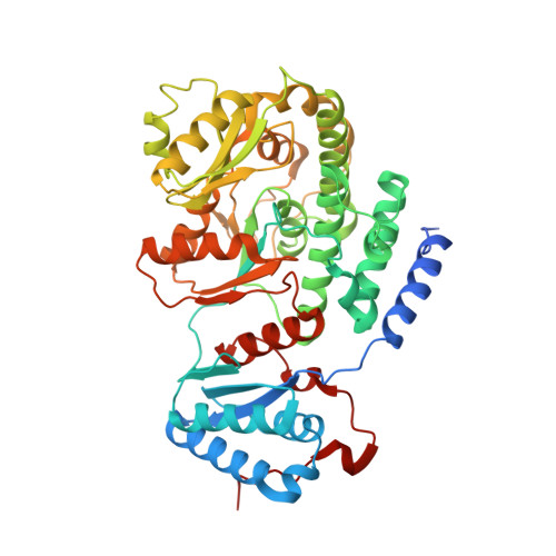
Biochemical and structural study of the atypical acyltransferase domain from the mycobacterial polyketide synthase pks13

Bergeret, F.Gavalda, S.Chalut, C.Malaga, W.Quemard, A.Pedelacq, J.D.Daffe, M.Guilhot, C.Mourey, L.Bon, C.

(2012) J Biological Chem 287: 33675-33690

  • DOI: https://doi.org/10.1074/jbc.M111.325639
  • Primary Citation Related Structures: 
    3TZW, 3TZX, 3TZY, 3TZZ

  • PubMed Abstract: 

    Pks13 is a type I polyketide synthase involved in the final biosynthesis step of mycolic acids, virulence factors, and essential components of the Mycobacterium tuberculosis envelope. Here, we report the biochemical and structural characterization of a 52-kDa fragment containing the acyltransferase domain of Pks13. This fragment retains the ability to load atypical extender units, unusually long chain acyl-CoA with a predilection for carboxylated substrates. High resolution crystal structures were determined for the apo, palmitoylated, and carboxypalmitoylated forms. Structural conservation with type I polyketide synthases and related fatty-acid synthases also extends to the interdomain connections. Subtle changes could be identified both in the active site and in the upstream and downstream linkers in line with the organization displayed by this singular polyketide synthase. More importantly, the crystallographic analysis illustrated for the first time how a long saturated chain can fit in the core structure of an acyltransferase domain through a dedicated channel. The structures also revealed the unexpected binding of a 12-mer peptide that might provide insight into domain-domain interaction.


  • Organizational Affiliation
    • Institut de Pharmacologie et de Biologie Structurale (IPBS), CNRS, Toulouse, France.

Macromolecule Content 

  • Total Structure Weight: 55.33 kDa 
  • Atom Count: 3,733 
  • Modeled Residue Count: 477 
  • Deposited Residue Count: 503 
  • Unique protein chains: 2

Macromolecules

Find similar proteins by:|  3D Structure
Entity ID: 1
MoleculeChains  Sequence LengthOrganismDetailsImage
Polyketide synthase PKS13491Mycobacterium tuberculosisMutation(s): 0 
Gene Names: Rv3800c
EC: 2.3.1
UniProt
Find proteins for O53579 (Mycobacterium tuberculosis (strain CDC 1551 / Oshkosh))
Explore O53579 
Go to UniProtKB:  O53579
Entity Groups
Sequence Clusters30% Identity50% Identity70% Identity90% Identity95% Identity100% Identity
UniProt GroupO53579
Sequence Annotations
Expand
Reference Sequence
Find similar proteins by:  Sequence   |   3D Structure  
Entity ID: 2
MoleculeChains  Sequence LengthOrganismDetailsImage
12-mer peptideB [auth D]12Escherichia coliMutation(s): 0 

Small Molecules

Ligands 2 Unique
IDChains Name / Formula / InChI Key2D Diagram3D Interactions
SO4

Query on SO4


Download:Ideal Coordinates CCD File
C [auth A]
D [auth A]
E [auth A]
F [auth A]
G [auth A]
C [auth A],
D [auth A],
E [auth A],
F [auth A],
G [auth A],
M [auth D]
SULFATE ION
O4 S
QAOWNCQODCNURD-UHFFFAOYSA-L
EDO

Query on EDO


Download:Ideal Coordinates CCD File
H [auth A],
I [auth A],
J [auth A],
K [auth A],
L [auth A]
1,2-ETHANEDIOL
C2 H6 O2
LYCAIKOWRPUZTN-UHFFFAOYSA-N

Experimental Data & Validation

Experimental Data

  • Method: X-RAY DIFFRACTION
  • Resolution: 2.60 Å
  • R-Value Free:  0.264 (Depositor), 0.253 (DCC) 
  • R-Value Work:  0.187 (Depositor), 0.186 (DCC) 
  • R-Value Observed: 0.190 (Depositor) 
Space Group: P 21 21 2
Unit Cell:
Length ( Å )Angle ( ˚ )
a = 108.659α = 90
b = 119.806β = 90
c = 50.162γ = 90
Software Package:
Software NamePurpose
DNAdata collection
PHASERphasing
REFMACrefinement
MOSFLMdata reduction
SCALAdata scaling

Structure Validation

View Full Validation Report


Entry History 

Deposition Data

Revision History  (Full details and data files)

  • Version 1.0: 2012-08-29
    Type: Initial release
  • Version 1.1: 2012-10-17
    Changes: Database references
  • Version 1.2: 2023-11-01
    Changes: Data collection, Database references, Derived calculations, Refinement description