3TZN | pdb_00003tzn

Crystal Structure of the Yersinia pestis Dihydropteroate synthase.


Experimental Data Snapshot

  • Method: X-RAY DIFFRACTION
  • Resolution: 2.08 Å
  • R-Value Free: 
    0.250 (Depositor), 0.250 (DCC) 
  • R-Value Work: 
    0.208 (Depositor), 0.209 (DCC) 
  • R-Value Observed: 
    0.210 (Depositor) 

wwPDB Validation 3D Report Full Report

Validation slider image for 3TZN

This is version 1.1 of the entry. See complete history

Literature

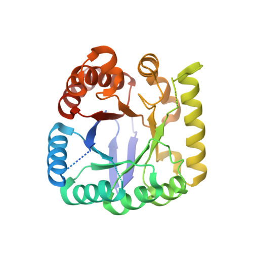
Catalysis and sulfa drug resistance in dihydropteroate synthase.

Yun, M.K.Wu, Y.Li, Z.Zhao, Y.Waddell, M.B.Ferreira, A.M.Lee, R.E.Bashford, D.White, S.W.

(2012) Science 335: 1110-1114

  • DOI: https://doi.org/10.1126/science.1214641
  • Primary Citation Related Structures: 
    3TYA, 3TYB, 3TYC, 3TYD, 3TYE, 3TYU, 3TYZ, 3TZF, 3TZN, 3V5O

  • PubMed Abstract: 

    The sulfonamide antibiotics inhibit dihydropteroate synthase (DHPS), a key enzyme in the folate pathway of bacteria and primitive eukaryotes. However, resistance mutations have severely compromised the usefulness of these drugs. We report structural, computational, and mutagenesis studies on the catalytic and resistance mechanisms of DHPS. By performing the enzyme-catalyzed reaction in crystalline DHPS, we have structurally characterized key intermediates along the reaction pathway. Results support an S(N)1 reaction mechanism via formation of a novel cationic pterin intermediate. We also show that two conserved loops generate a substructure during catalysis that creates a specific binding pocket for p-aminobenzoic acid, one of the two DHPS substrates. This substructure, together with the pterin-binding pocket, explains the roles of the conserved active-site residues and reveals how sulfonamide resistance arises.


  • Organizational Affiliation
    • Department of Structural Biology, St. Jude Children's Research Hospital, Memphis, TN 38105, USA.

Macromolecule Content 

  • Total Structure Weight: 60.64 kDa 
  • Atom Count: 3,561 
  • Modeled Residue Count: 455 
  • Deposited Residue Count: 560 
  • Unique protein chains: 1

Macromolecules

Find similar proteins by:|  3D Structure
Entity ID: 1
MoleculeChains  Sequence LengthOrganismDetailsImage
7,8-dihydropteroate synthase
A, B
280Yersinia pestisMutation(s): 0 
Gene Names: dhpSfolPy0683YPO3501YP_0582
EC: 2.5.1.15
UniProt
Find proteins for A0A2S9PLG4 (Yersinia pestis)
Explore A0A2S9PLG4 
Go to UniProtKB:  A0A2S9PLG4
Entity Groups
Sequence Clusters30% Identity50% Identity70% Identity90% Identity95% Identity100% Identity
UniProt GroupA0A2S9PLG4
Sequence Annotations
Expand
Reference Sequence

Experimental Data & Validation

Experimental Data

  • Method: X-RAY DIFFRACTION
  • Resolution: 2.08 Å
  • R-Value Free:  0.250 (Depositor), 0.250 (DCC) 
  • R-Value Work:  0.208 (Depositor), 0.209 (DCC) 
  • R-Value Observed: 0.210 (Depositor) 
Space Group: P 1 21 1
Unit Cell:
Length ( Å )Angle ( ˚ )
a = 71.429α = 90
b = 49.936β = 90.24
c = 75.512γ = 90
Software Package:
Software NamePurpose
SCALEPACKdata scaling
PHENIXrefinement
PDB_EXTRACTdata extraction
MAR345dtbdata collection
HKL-2000data reduction
PHENIXphasing

Structure Validation

View Full Validation Report


Entry History 

Deposition Data

  • Released Date: 2012-03-14 
  • Deposition Author(s): Wu, Y.

Revision History  (Full details and data files)

  • Version 1.0: 2012-03-14
    Type: Initial release
  • Version 1.1: 2024-02-28
    Changes: Data collection, Database references