Protein camouflage in cytochrome c-calixarene complexes.
McGovern, R.E., Fernandes, H., Khan, A.R., Power, N.P., Crowley, P.B.(2012) Nat Chem 4: 527-533
- PubMed: 22717436 Search on PubMed
- DOI: https://doi.org/10.1038/nchem.1342
- Primary Citation Related Structures:
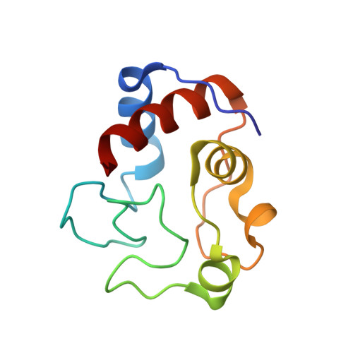
3TYI - PubMed Abstract:
Small molecules that recognize protein surfaces are important tools for modifying protein interaction properties. Since the 1980s, several thousand studies concerning calixarenes and host-guest interactions have been published. Although there is growing interest in protein-calixarene interactions, only limited structural information has been available to date. We now report the crystal structure of a protein-calixarene complex. The water-soluble p-sulfonatocalix[4]arene is shown to bind the lysine-rich cytochrome c at three different sites. Binding curves obtained from NMR titrations reveal an interaction process that involves two or more binding sites. Together, the data indicate a dynamic complex in which the calixarene explores the surface of cytochrome c. In addition to providing valuable information on protein recognition, the data also indicate that the calixarene is a mediator of protein-protein interactions, with potential applications in generating assemblies and promoting crystallization.
- School of Chemistry, National University of Ireland Galway, University Road, Galway, Ireland.
Organizational Affiliation: