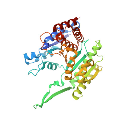
3TY3

Crystal structure of homoisocitrate dehydrogenase from Schizosaccharomyces pombe bound to glycyl-glycyl-glycine


Experimental Data Snapshot

  • Method: X-RAY DIFFRACTION
  • Resolution: 1.85 Å
  • R-Value Free: 0.230 
  • R-Value Work: 0.193 
  • R-Value Observed: 0.195 

Starting Model: experimental
View more details

wwPDB Validation   3D Report Full Report


Ligand Structure Quality Assessment 


This is version 1.4 of the entry. See complete history


Literature

Crystal structure of homoisocitrate dehydrogenase from Schizosaccharomyces pombe.

Bulfer, S.L.Hendershot, J.M.Trievel, R.C.

(2012) Proteins 80: 661-666

  • DOI: https://doi.org/10.1002/prot.23231
  • Primary Citation of Related Structures:  
    3TY3, 3TY4

  • PubMed Abstract: 

    Homoisocitrate dehydrogenase (HICDH) catalyzes the conversion of homoisocitrate to 2-oxoadipate, the third enzymatic step in the α-aminoadipate pathway by which lysine is synthesized in fungi and certain archaebacteria. This enzyme represents a potential target for anti-fungal drug design. Here, we describe the first crystal structures of a fungal HICDH, including structures of an apoenzyme and a binary complex with a glycine tri-peptide. The structures illustrate the homology of HICDH with other β-hydroxyacid oxidative decarboxylases and reveal key differences with the active site of Thermus thermophilus HICDH that provide insights into the differences in substrate specificity of these enzymes.


  • Organizational Affiliation

    Department of Biological Chemistry, University of Michigan, Ann Arbor, Michigan 48109.


Macromolecules
Find similar proteins by:  (by identity cutoff)  |  3D Structure
Entity ID: 1
MoleculeChains Sequence LengthOrganismDetailsImage
Probable homoisocitrate dehydrogenase
A, B
366Schizosaccharomyces pombe 972h-Mutation(s): 0 
Gene Names: lys12SPAC31G5.04
EC: 1.1.1.87
UniProt
Find proteins for O14104 (Schizosaccharomyces pombe (strain 972 / ATCC 24843))
Explore O14104 
Go to UniProtKB:  O14104
Entity Groups  
Sequence Clusters30% Identity50% Identity70% Identity90% Identity95% Identity100% Identity
UniProt GroupO14104
Sequence Annotations
Expand
  • Reference Sequence
Experimental Data & Validation

Experimental Data

  • Method: X-RAY DIFFRACTION
  • Resolution: 1.85 Å
  • R-Value Free: 0.230 
  • R-Value Work: 0.193 
  • R-Value Observed: 0.195 
  • Space Group: P 1 21 1
Unit Cell:
Length ( Å )Angle ( ˚ )
a = 53.243α = 90
b = 92.854β = 106.71
c = 75.474γ = 90
Software Package:
Software NamePurpose
DENZOdata reduction
SCALEPACKdata scaling
REFMACrefinement
PDB_EXTRACTdata extraction
HKL-2000data collection
HKL-2000data reduction
HKL-2000data scaling
MOLREPphasing

Structure Validation

View Full Validation Report


Ligand Structure Quality Assessment 


Entry History 

Deposition Data

Revision History  (Full details and data files)

  • Version 1.0: 2011-11-09
    Type: Initial release
  • Version 1.1: 2011-12-14
    Changes: Database references
  • Version 1.2: 2013-11-27
    Changes: Database references
  • Version 1.3: 2017-11-08
    Changes: Refinement description
  • Version 1.4: 2023-09-13
    Changes: Data collection, Database references, Derived calculations, Refinement description