3TVY | pdb_00003tvy

Structural Analysis of Adhesive Tip pilin, GBS104 from Group B Streptococcus agalactiae


Experimental Data Snapshot

  • Method: X-RAY DIFFRACTION
  • Resolution: 2.00 Å
  • R-Value Free: 
    0.251 (Depositor), 0.229 (DCC) 
  • R-Value Work: 
    0.210 (Depositor) 
  • R-Value Observed: 
    0.212 (Depositor) 

wwPDB Validation 3D Report Full Report

Validation slider image for 3TVY

This is version 1.3 of the entry. See complete history

Literature

Structure of Streptococcus agalactiae tip pilin GBS104: a model for GBS pili assembly and host interactions.

Krishnan, V.Dwivedi, P.Kim, B.J.Samal, A.Macon, K.Ma, X.Mishra, A.Doran, K.S.Ton-That, H.Narayana, S.V.

(2013) Acta Crystallogr D Biol Crystallogr 69: 1073-1089

  • DOI: https://doi.org/10.1107/S0907444913004642
  • Primary Citation Related Structures: 
    3TVY, 3TW0, 3TXA

  • PubMed Abstract: 

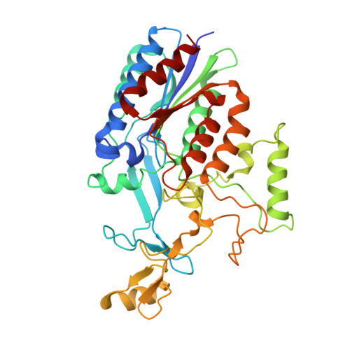
    The crystal structure of a 75 kDa central fragment of GBS104, a tip pilin from the 2063V/R strain of Streptococcus agalactiae (group B streptococcus; GBS), is reported. In addition, a homology model of the remaining two domains of GBS104 was built and a model of full-length GBS104 was generated by combining the homology model (the N1 and N4 domains) and the crystal structure of the 75 kDa fragment (the N2 and N3 domains). This rod-shaped GBS104 model is constructed of three IgG-like domains (the N1, N2 and N4 domains) and one vWFA-like domain (the N3 domain). The N1 and N2 domains of GBS104 are assembled with distinct and remote segments contributed by the N- and C-termini. The metal-binding site in the N3 domain of GBS104 is in the closed/low-affinity conformation. Interestingly, this domain hosts two long arms that project away from the metal-binding site. Using site-directed mutagenesis, two cysteine residues that lock the N3 domain of GBS104 into the open/high-affinity conformation were introduced. Both wild-type and disulfide-locked recombinant proteins were tested for binding to extracellular matrix proteins such as collagen, fibronectin, fibrinogen and laminin, and an increase in fibronectin binding affinity was identified for the disulfide-locked N3 domain, suggesting that induced conformational changes may play a possible role in receptor binding.


  • Organizational Affiliation
    • UNESCO Regional Centre for Biotechnology (RCB), Gurgaon 122 016, Haryana, India.

Macromolecule Content 

  • Total Structure Weight: 42.24 kDa 
  • Atom Count: 3,150 
  • Modeled Residue Count: 365 
  • Deposited Residue Count: 373 
  • Unique protein chains: 1

Macromolecules

Find similar proteins by:|  3D Structure
Entity ID: 1
MoleculeChains  Sequence LengthOrganismDetailsImage
Cell wall surface anchor family protein373Streptococcus agalactiae serogroup VMutation(s): 0 
Gene Names: SAG0649
UniProt
Find proteins for Q8E0S5 (Streptococcus agalactiae serotype V (strain ATCC BAA-611 / 2603 V/R))
Explore Q8E0S5 
Go to UniProtKB:  Q8E0S5
Entity Groups
Sequence Clusters30% Identity50% Identity70% Identity90% Identity95% Identity100% Identity
UniProt GroupQ8E0S5
Sequence Annotations
Expand
Reference Sequence

Small Molecules

Ligands 2 Unique
IDChains Name / Formula / InChI Key2D Diagram3D Interactions
EPE

Query on EPE


Download:Ideal Coordinates CCD File
B [auth A]4-(2-HYDROXYETHYL)-1-PIPERAZINE ETHANESULFONIC ACID
C8 H18 N2 O4 S
JKMHFZQWWAIEOD-UHFFFAOYSA-N
MG

Query on MG


Download:Ideal Coordinates CCD File
C [auth A]MAGNESIUM ION
Mg
JLVVSXFLKOJNIY-UHFFFAOYSA-N

Experimental Data & Validation

Experimental Data

  • Method: X-RAY DIFFRACTION
  • Resolution: 2.00 Å
  • R-Value Free:  0.251 (Depositor), 0.229 (DCC) 
  • R-Value Work:  0.210 (Depositor) 
  • R-Value Observed: 0.212 (Depositor) 
Space Group: P 21 21 2
Unit Cell:
Length ( Å )Angle ( ˚ )
a = 74.749α = 90
b = 101.29β = 90
c = 52.665γ = 90
Software Package:
Software NamePurpose
StructureStudiodata collection
PHENIXmodel building
REFMACrefinement
d*TREKdata reduction
d*TREKdata scaling
PHENIXphasing

Structure Validation

View Full Validation Report


Entry History 

Deposition Data

Revision History  (Full details and data files)

  • Version 1.0: 2013-03-27
    Type: Initial release
  • Version 1.1: 2013-05-29
    Changes: Database references
  • Version 1.2: 2013-06-19
    Changes: Database references
  • Version 1.3: 2024-02-28
    Changes: Data collection, Database references, Derived calculations