3TUH | pdb_00003tuh

Crystal Structure of the N-terminal domain of an HSP90 in the presence of an the inhibitor ganetespib


Experimental Data Snapshot

  • Method: X-RAY DIFFRACTION
  • Resolution: 1.80 Å
  • R-Value Free: 
    0.212 (Depositor), 0.210 (DCC) 
  • R-Value Work: 
    0.172 (Depositor), 0.170 (DCC) 

wwPDB Validation   3D Report Full Report


Ligand Structure Quality Assessment 

Created with Raphaël 2.3.0Worse 01 BetterLigand structure goodness of fit to experimental dataBest fitted TUHClick on this verticalbar to view details

This is version 1.3 of the entry. See complete history


Literature

Crystal Structure of the N-terminal domain of an HSP90 in the presence of an the inhibitor ganetespib

Ying, W.

To be published.

Macromolecules
Find similar proteins by:  (by identity cutoff)  |  3D Structure
Entity ID: 1
MoleculeChains Sequence LengthOrganismDetailsImage
Heat shock protein HSP 90-alpha
A, B
209Homo sapiensMutation(s): 0 
Gene Names: HSP90AA1HSP90AHSPC1HSPCA
EC: 3.6.4.10
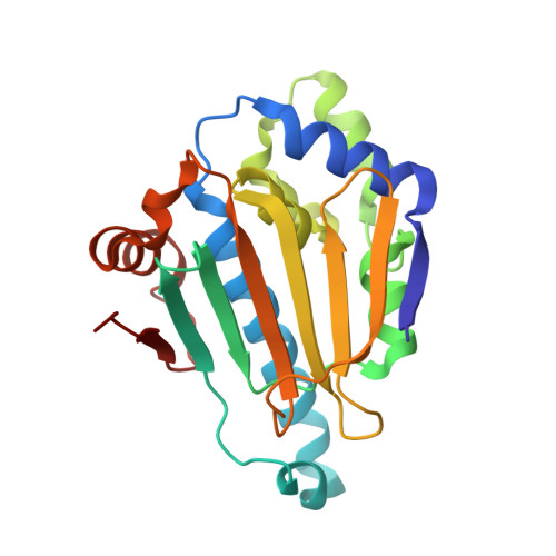
UniProt & NIH Common Fund Data Resources
Find proteins for P07900 (Homo sapiens)
Explore P07900 
Go to UniProtKB:  P07900
PHAROS:  P07900
GTEx:  ENSG00000080824 
Entity Groups  
Sequence Clusters30% Identity50% Identity70% Identity90% Identity95% Identity100% Identity
UniProt GroupP07900
Sequence Annotations
Expand
  • Reference Sequence
Small Molecules
Ligands 1 Unique
IDChains Name / Formula / InChI Key2D Diagram3D Interactions
TUH
Query on TUH

Download Ideal Coordinates CCD File 
C [auth A],
D [auth B]
5-[2,4-dihydroxy-5-(propan-2-yl)phenyl]-4-(1-methyl-1H-indol-5-yl)-2,4-dihydro-3H-1,2,4-triazol-3-one
C20 H20 N4 O3
RVAQIUULWULRNW-UHFFFAOYSA-N
Binding Affinity Annotations 
IDSourceBinding Affinity
TUH BindingDB:  3TUH Kd: 24 (nM) from 1 assay(s)
IC50: min: 6, max: 5390 (nM) from 4 assay(s)
Experimental Data & Validation

Experimental Data

  • Method: X-RAY DIFFRACTION
  • Resolution: 1.80 Å
  • R-Value Free:  0.212 (Depositor), 0.210 (DCC) 
  • R-Value Work:  0.172 (Depositor), 0.170 (DCC) 
Space Group: P 1 21 1
Unit Cell:
Length ( Å )Angle ( ˚ )
a = 56.362α = 90
b = 88.878β = 99.41
c = 56.498γ = 90
Software Package:
Software NamePurpose
REFMACrefinement

Structure Validation

View Full Validation Report


Ligand Structure Quality Assessment 

Created with Raphaël 2.3.0Worse 01 BetterLigand structure goodness of fit to experimental dataBest fitted TUHClick on this verticalbar to view details

Entry History 

Deposition Data

  • Released Date: 2012-09-19 
  • Deposition Author(s): Ying, W.

Revision History  (Full details and data files)

  • Version 1.0: 2012-09-19
    Type: Initial release
  • Version 1.1: 2018-03-07
    Changes: Data collection
  • Version 1.2: 2021-11-17
    Changes: Database references, Derived calculations, Structure summary
  • Version 1.3: 2024-02-28
    Changes: Data collection