3TTC

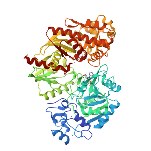
Crystal structure of E. coli HypF with ADP and carbamoyl phosphate


Experimental Data Snapshot

  • Method: X-RAY DIFFRACTION
  • Resolution: 1.86 Å
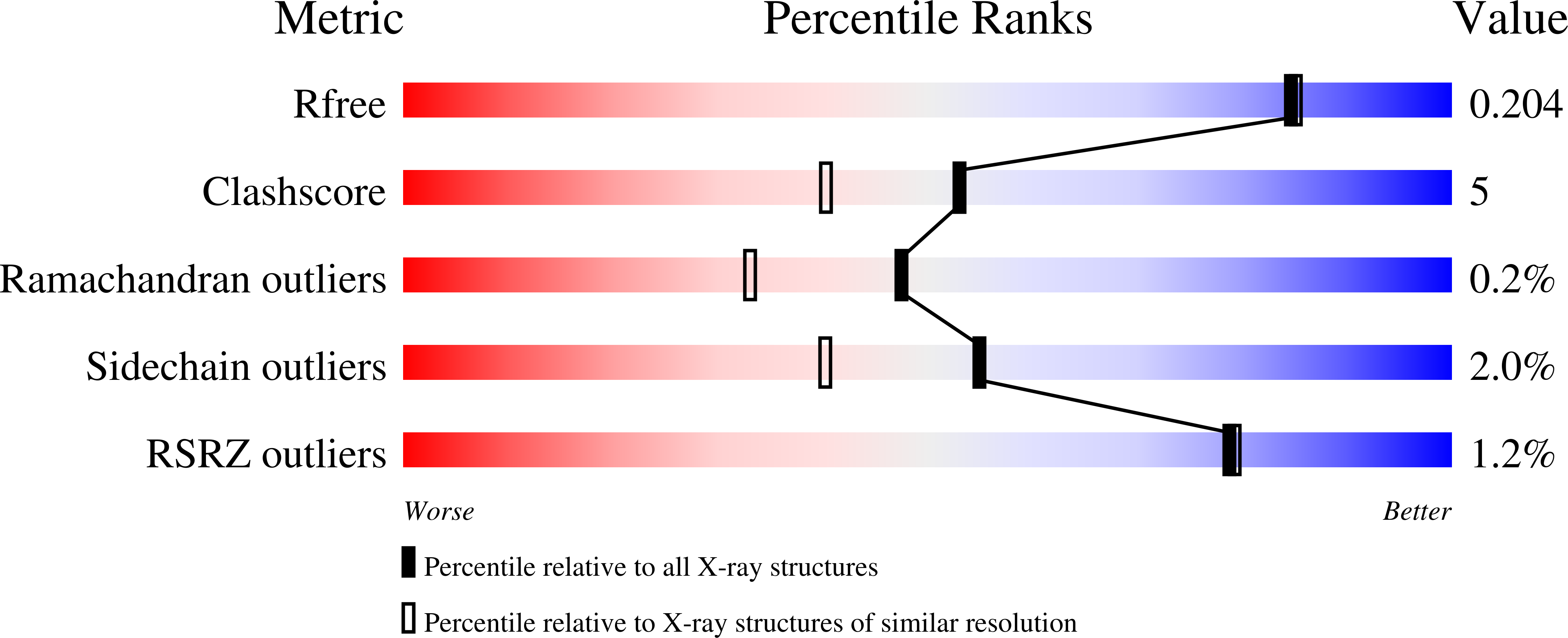
  • R-Value Free: 0.202 
  • R-Value Work: 0.159 
  • R-Value Observed: 0.161 

wwPDB Validation   3D Report Full Report


Ligand Structure Quality Assessment 


This is version 1.1 of the entry. See complete history


Literature

Structure of Hydrogenase Maturation Protein HypF with Reaction Intermediates Shows Two Active Sites.

Petkun, S.Shi, R.Li, Y.Asinas, A.Munger, C.Zhang, L.Waclawek, M.Soboh, B.Sawers, R.G.Cygler, M.

(2011) Structure 19: 1773-1783

  • DOI: https://doi.org/10.1016/j.str.2011.09.023
  • Primary Citation of Related Structures:  
    3TSP, 3TSQ, 3TSU, 3TTC, 3TTD, 3TTF

  • PubMed Abstract: 

    [NiFe]-hydrogenases are multimeric proteins. The large subunit contains the NiFe(CN)(2)CO bimetallic active center and the small subunit contains Fe-S clusters. Biosynthesis and assembly of the NiFe(CN)(2)CO active center requires six Hyp accessory proteins. The synthesis of the CN(-) ligands is catalyzed by the combined actions of HypF and HypE using carbamoylphosphate as a substrate. We report the structure of Escherichia coli HypF(92-750) lacking the N-terminal acylphosphatase domain. HypF(92-750) comprises the novel Zn-finger domain, the nucleotide-binding YrdC-like domain, and the Kae1-like universal domain, also binding a nucleotide and a Zn(2+) ion. The two nucleotide-binding sites are sequestered in an internal cavity, facing each other and separated by ∼14 Å. The YrdC-like domain converts carbamoyl moiety to a carbamoyl adenylate intermediate, which is channeled to the Kae1-like domain. Mutations within either nucleotide-binding site compromise hydrogenase maturation but do not affect the carbamoylphosphate phosphatase activity.


  • Organizational Affiliation

    Department of Biochemistry, McGill University, Montreal, QC H3G 1Y6, Canada.


Macromolecules
Find similar proteins by:  (by identity cutoff)  |  3D Structure
Entity ID: 1
MoleculeChains Sequence LengthOrganismDetailsImage
Transcriptional regulatory protein657Escherichia coli O157:H7Mutation(s): 0 
Gene Names: ECs3568hypf
EC: 2.1.3 (PDB Primary Data), 6.2 (UniProt)
UniProt
Find proteins for A0A0H3JHT3 (Escherichia coli O157:H7)
Explore A0A0H3JHT3 
Go to UniProtKB:  A0A0H3JHT3
Entity Groups  
Sequence Clusters30% Identity50% Identity70% Identity90% Identity95% Identity100% Identity
UniProt GroupA0A0H3JHT3
Sequence Annotations
Expand
  • Reference Sequence
Experimental Data & Validation

Experimental Data

  • Method: X-RAY DIFFRACTION
  • Resolution: 1.86 Å
  • R-Value Free: 0.202 
  • R-Value Work: 0.159 
  • R-Value Observed: 0.161 
  • Space Group: P 21 21 21
Unit Cell:
Length ( Å )Angle ( ˚ )
a = 46.36α = 90
b = 77.913β = 90
c = 200.488γ = 90
Software Package:
Software NamePurpose
REFMACrefinement
PDB_EXTRACTdata extraction
MxDCdata collection

Structure Validation

View Full Validation Report


Ligand Structure Quality Assessment 


Entry History 

Deposition Data

Revision History  (Full details and data files)

  • Version 1.0: 2011-12-28
    Type: Initial release
  • Version 1.1: 2024-02-28
    Changes: Data collection, Database references, Derived calculations