3TSR | pdb_00003tsr

X-ray structure of mouse ribonuclease inhibitor complexed with mouse ribonuclease 1


Experimental Data Snapshot

  • Method: X-RAY DIFFRACTION
  • Resolution: 2.20 Å
  • R-Value Free: 
    0.233 (Depositor), 0.230 (DCC) 
  • R-Value Work: 
    0.182 (Depositor), 0.179 (DCC) 
  • R-Value Observed: 
    0.183 (Depositor) 

wwPDB Validation 3D Report Full Report

Validation slider image for 3TSR

This is version 1.5 of the entry. See complete history

Literature

Functional evolution of ribonuclease inhibitor: insights from birds and reptiles.

Lomax, J.E.Bianchetti, C.M.Chang, A.Phillips, G.N.Fox, B.G.Raines, R.T.

(2014) J Mol Biology 426: 3041-3056

  • DOI: https://doi.org/10.1016/j.jmb.2014.06.007
  • Primary Citation Related Structures: 
    3TSR, 4PEQ, 4PER

  • PubMed Abstract: 

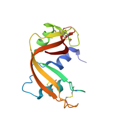
    Ribonuclease inhibitor (RI) is a conserved protein of the mammalian cytosol. RI binds with high affinity to diverse secretory ribonucleases (RNases) and inhibits their enzymatic activity. Although secretory RNases are found in all vertebrates, the existence of a non-mammalian RI has been uncertain. Here, we report on the identification and characterization of RI homologs from chicken and anole lizard. These proteins bind to RNases from multiple species but exhibit much greater affinity for their cognate RNases than for mammalian RNases. To reveal the basis for this differential affinity, we determined the crystal structure of mouse, bovine, and chicken RI·RNase complexes to a resolution of 2.20, 2.21, and 1.92Å, respectively. A combination of structural, computational, and bioinformatic analyses enabled the identification of two residues that appear to contribute to the differential affinity for RNases. We also found marked differences in oxidative instability between mammalian and non-mammalian RIs, indicating evolution toward greater oxygen sensitivity in RIs from mammalian species. Taken together, our results illuminate the structural and functional evolution of RI, along with its dynamic role in vertebrate biology.


  • Organizational Affiliation
    • Graduate Program in Cellular and Molecular Biology, University of Wisconsin-Madison, Madison, WI 53706, USA.

Macromolecule Content 

  • Total Structure Weight: 257.56 kDa 
  • Atom Count: 18,531 
  • Modeled Residue Count: 2,305 
  • Deposited Residue Count: 2,328 
  • Unique protein chains: 2

Macromolecules

Find similar proteins by:|  3D Structure
Entity ID: 1
MoleculeChains  Sequence LengthOrganismDetailsImage
Ribonuclease pancreatic
A, B, C, D
125Mus musculusMutation(s): 0 
Gene Names: Rib-1Rib1Rnase1Rns1
EC: 3.1.27.5 (PDB Primary Data), 4.6.1.18 (UniProt)
UniProt & NIH Common Fund Data Resources
Find proteins for P00683 (Mus musculus)
Explore P00683 
Go to UniProtKB:  P00683
IMPC:  MGI:97919
Entity Groups
Sequence Clusters30% Identity50% Identity70% Identity90% Identity95% Identity100% Identity
UniProt GroupP00683
Sequence Annotations
Expand
Reference Sequence
Find similar proteins by:|  3D Structure
Entity ID: 2
MoleculeChains  Sequence LengthOrganismDetailsImage
Ribonuclease inhibitor
E, F, G, H
457Mus musculusMutation(s): 0 
Gene Names: RnhRnh1
UniProt
Find proteins for Q91VI7 (Mus musculus)
Explore Q91VI7 
Go to UniProtKB:  Q91VI7
Entity Groups
Sequence Clusters30% Identity50% Identity70% Identity90% Identity95% Identity100% Identity
UniProt GroupQ91VI7
Sequence Annotations
Expand
Reference Sequence

Small Molecules

Ligands 2 Unique
IDChains Name / Formula / InChI Key2D Diagram3D Interactions
PEG

Query on PEG


Download:Ideal Coordinates CCD File
J [auth E]DI(HYDROXYETHYL)ETHER
C4 H10 O3
MTHSVFCYNBDYFN-UHFFFAOYSA-N
EDO

Query on EDO


Download:Ideal Coordinates CCD File
I [auth A]
K [auth E]
L [auth E]
M [auth F]
N [auth F]
I [auth A],
K [auth E],
L [auth E],
M [auth F],
N [auth F],
O [auth F],
P [auth G],
Q [auth G],
R [auth G],
S [auth G],
T [auth H],
U [auth H],
V [auth H]
1,2-ETHANEDIOL
C2 H6 O2
LYCAIKOWRPUZTN-UHFFFAOYSA-N

Experimental Data & Validation

Experimental Data

  • Method: X-RAY DIFFRACTION
  • Resolution: 2.20 Å
  • R-Value Free:  0.233 (Depositor), 0.230 (DCC) 
  • R-Value Work:  0.182 (Depositor), 0.179 (DCC) 
  • R-Value Observed: 0.183 (Depositor) 
Space Group: P 1 21 1
Unit Cell:
Length ( Å )Angle ( ˚ )
a = 72.396α = 90
b = 125.341β = 94.72
c = 123.064γ = 90
Software Package:
Software NamePurpose
DENZOdata reduction
SCALEPACKdata scaling
PHASERphasing
PHENIXrefinement
PDB_EXTRACTdata extraction
HKL-2000data collection
HKL-2000data reduction
HKL-2000data scaling

Structure Validation

View Full Validation Report


Entry History 

Deposition Data

Revision History  (Full details and data files)

  • Version 1.0: 2012-09-19
    Type: Initial release
  • Version 1.1: 2014-06-18
    Changes: Database references
  • Version 1.2: 2014-07-02
    Changes: Database references
  • Version 1.3: 2014-08-20
    Changes: Database references
  • Version 1.4: 2017-11-08
    Changes: Refinement description
  • Version 1.5: 2024-10-16
    Changes: Data collection, Database references, Derived calculations, Structure summary