3TRY | pdb_00003try

Crystal structure of racemic villin headpiece subdomain in space group I-4c2


Experimental Data Snapshot

  • Method: X-RAY DIFFRACTION
  • Resolution: 2.30 Å
  • R-Value Free: 
    0.230 (Depositor), 0.228 (DCC) 
  • R-Value Work: 
    0.200 (Depositor), 0.198 (DCC) 
  • R-Value Observed: 
    0.202 (Depositor) 

Starting Model: experimental
View more details

wwPDB Validation 3D Report Full Report

Validation slider image for 3TRY

This is version 1.5 of the entry. See complete history

Literature

Quasiracemic crystallization as a tool to assess the accommodation of noncanonical residues in nativelike protein conformations.

Mortenson, D.E.Satyshur, K.A.Guzei, I.A.Forest, K.T.Gellman, S.H.

(2012) J Am Chem Soc 134: 2473-2476

  • DOI: https://doi.org/10.1021/ja210045s
  • Primary Citation Related Structures: 
    3TJW, 3TRV, 3TRW, 3TRY

  • PubMed Abstract: 

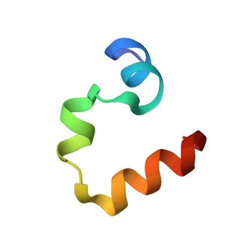
    Quasiracemic crystallization has been used to obtain high-resolution structures of two variants of the villin headpiece subdomain (VHP) that contain a pentafluorophenylalanine (F(5)Phe) residue in the hydrophobic core. In each case, the crystal contained the variant constructed from l-amino acids and the native sequence constructed from d-amino acids. We were motivated to undertake these studies by reports that racemic proteins crystallize more readily than homochiral forms and the prospect that quasiracemic crystallization would enable us to determine whether a polypeptide containing a noncanonical residue can closely mimic the tertiary structure of the native sequence. The results suggest that quasiracemic crystallization may prove to be generally useful for assessing mimicry of naturally evolved protein folding patterns by polypeptides that contain unnatural side-chain or backbone subunits.


  • Organizational Affiliation
    • Department of Chemistry, University of Wisconsin, Madison, Wisconsin 53706, USA.

Macromolecule Content 

  • Total Structure Weight: 4.18 kDa 
  • Atom Count: 285 
  • Modeled Residue Count: 34 
  • Deposited Residue Count: 35 
  • Unique protein chains: 1

Macromolecules

Find similar proteins by:|  3D Structure
Entity ID: 1
MoleculeChains  Sequence LengthOrganismDetailsImage
D-Villin-135Gallus gallusMutation(s): 1 
UniProt
Find proteins for P02640 (Gallus gallus)
Explore P02640 
Go to UniProtKB:  P02640
Entity Groups
Sequence Clusters30% Identity50% Identity70% Identity90% Identity95% Identity100% Identity
UniProt GroupP02640
Sequence Annotations
Expand
Reference Sequence

Small Molecules

Ligands 1 Unique
IDChains Name / Formula / InChI Key2D Diagram3D Interactions
SO4

Query on SO4


Download:Ideal Coordinates CCD File
B [auth A]SULFATE ION
O4 S
QAOWNCQODCNURD-UHFFFAOYSA-L

Experimental Data & Validation

Experimental Data

  • Method: X-RAY DIFFRACTION
  • Resolution: 2.30 Å
  • R-Value Free:  0.230 (Depositor), 0.228 (DCC) 
  • R-Value Work:  0.200 (Depositor), 0.198 (DCC) 
  • R-Value Observed: 0.202 (Depositor) 
Space Group: I -4 c 2
Unit Cell:
Length ( Å )Angle ( ˚ )
a = 63.128α = 90
b = 63.128β = 90
c = 39.135γ = 90
Software Package:
Software NamePurpose
SAINTdata reduction
REFMACrefinement
PDB_EXTRACTdata extraction
PROTEUM PLUSdata reduction
SAINTdata scaling
PHASERphasing

Structure Validation

View Full Validation Report


Entry History 

Deposition Data

Revision History  (Full details and data files)

  • Version 1.0: 2012-01-25
    Type: Initial release
  • Version 1.1: 2012-02-08
    Changes: Database references
  • Version 1.2: 2012-02-22
    Changes: Database references
  • Version 1.3: 2023-09-13
    Changes: Data collection, Database references, Derived calculations, Refinement description
  • Version 1.4: 2023-12-06
    Changes: Data collection
  • Version 1.5: 2024-10-30
    Changes: Structure summary