3TO4 | pdb_00003to4

Structure of mouse Valpha14Vbeta2-mouseCD1d-alpha-Galactosylceramide


Experimental Data Snapshot

  • Method: X-RAY DIFFRACTION
  • Resolution: 3.10 Å
  • R-Value Free: 
    0.268 (Depositor), 0.261 (DCC) 
  • R-Value Work: 
    0.217 (Depositor), 0.214 (DCC) 
  • R-Value Observed: 
    0.220 (Depositor) 

Starting Model: experimental
View more details

wwPDB Validation 3D Report Full Report

Validation slider image for 3TO4

Ligand Structure Quality Assessment 


This is version 2.2 of the entry. See complete history

Literature

Vbeta2 natural killer T cell antigen receptor-mediated recognition of CD1d-glycolipid antigen.

Patel, O.Pellicci, D.G.Uldrich, A.P.Sullivan, L.C.Bhati, M.McKnight, M.Richardson, S.K.Howell, A.R.Mallevaey, T.Zhang, J.Bedel, R.Besra, G.S.Brooks, A.G.Kjer-Nielsen, L.McCluskey, J.Porcelli, S.A.Gapin, L.Rossjohn, J.Godfrey, D.I.

(2011) Proc Natl Acad Sci U S A 108: 19007-19012

  • DOI: https://doi.org/10.1073/pnas.1109066108
  • Primary Citation Related Structures: 
    3TO4

  • PubMed Abstract: 

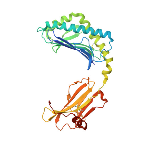
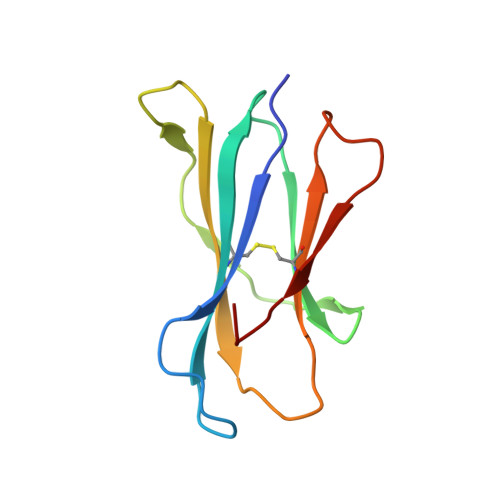
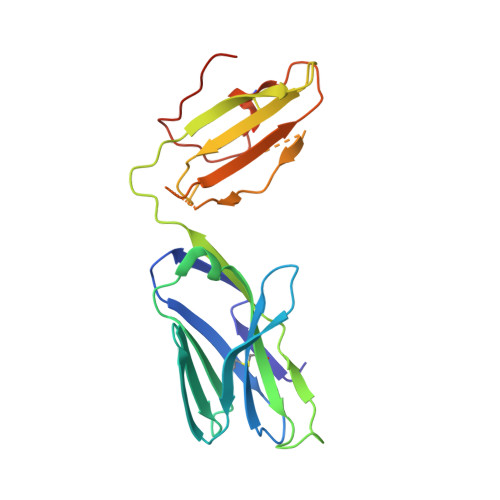
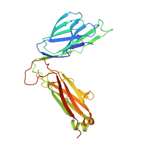
    Natural killer T cell antigen receptors (NKT TCRs) recognize lipid-based antigens (Ags) presented by CD1d. Although the TCR α-chain is invariant, NKT TCR Vβ exhibits greater diversity, with one (Vβ11) and three (Vβ8, Vβ7, and Vβ2) Vβ chains in humans and mice, respectively. With the exception of the Vβ2 NKT TCR, NKT TCRs possess canonical tyrosine residues within complementarity determining region (CDR) 2β that are critical for CD1d binding. Thus, how Vβ2 NKT TCR docks with CD1d-Ag was unclear. Despite the absence of the CDR2β-encoded tyrosine residues, we show that the Vβ2 NKT TCR engaged CD1d-Ag in a similar manner and with a comparable affinity and energetic footprint to the manner observed for the Vβ8.2 and Vβ7 NKT TCRs. Accordingly, the germline-encoded regions of the TCR β-chain do not exclusively dictate the innate NKT TCR-CD1d-Ag docking mode. Nevertheless, clear fine specificity differences for the CD1d-Ag existed between the Vβ2 NKT TCR and the Vβ8.2 and Vβ7 NKT TCRs, with the Vβ2 NKT TCR exhibiting greater sensitivity to modifications to the glycolipid Ag. Furthermore, within the Vβ2 NKT TCR-CD1d-αGalCer complex, the CDR2β loop mediated fewer contacts with CD1d, whereas the CDR1β and CDR3β loops contacted CD1d to a much greater extent compared with most Vβ11, Vβ8.2, and Vβ7 NKT TCRs. Accordingly, there is a greater interplay between the germline- and nongermline-encoded loops within the TCR β-chain of the Vβ2 NKT TCR that enables CD1d-Ag ligation.


  • Organizational Affiliation
    • Protein Crystallography Unit, Australian Research Council Centre of Excellence in Structural and Functional Microbial Genomics, Department of Biochemistry and Molecular Biology, School of Biomedical Sciences, Monash University, Clayton, Victoria 3800, Australia.

Macromolecule Content 

  • Total Structure Weight: 100.18 kDa 
  • Atom Count: 6,496 
  • Modeled Residue Count: 820 
  • Deposited Residue Count: 866 
  • Unique protein chains: 4

Macromolecules

Find similar proteins by:|  3D Structure
Entity ID: 1
MoleculeChains  Sequence LengthOrganismDetailsImage
Antigen-presenting glycoprotein CD1d1302Mus musculusMutation(s): 0 
Gene Names: Cd1d1Cd1.1
UniProt & NIH Common Fund Data Resources
Find proteins for P11609 (Mus musculus)
Explore P11609 
Go to UniProtKB:  P11609
IMPC:  MGI:107674
Entity Groups
Sequence Clusters30% Identity50% Identity70% Identity90% Identity95% Identity100% Identity
UniProt GroupP11609
Glycosylation
Glycosylation Sites: 3Go to GlyGen: P11609-1
Sequence Annotations
Expand
Reference Sequence
Find similar proteins by:|  3D Structure
Entity ID: 2
MoleculeChains  Sequence LengthOrganismDetailsImage
Beta-2 microglobulin99Mus musculusMutation(s): 0 
Gene Names: B2mRP23-34E24.5-001mCG_11606
UniProt & NIH Common Fund Data Resources
Find proteins for P01887 (Mus musculus)
Explore P01887 
Go to UniProtKB:  P01887
IMPC:  MGI:88127
Entity Groups
Sequence Clusters30% Identity50% Identity70% Identity90% Identity95% Identity100% Identity
UniProt GroupP01887
Sequence Annotations
Expand
Reference Sequence
Find similar proteins by:|  3D Structure
Entity ID: 3
MoleculeChains  Sequence LengthOrganismDetailsImage
NKT Valpha14 (MOUSE VARIABLE DOMAIN, HUMAN CONSTANT DOMAIN)212Homo sapiensMus musculus
This entity is chimeric
Mutation(s): 0 
Find similar proteins by:|  3D Structure
Entity ID: 4
MoleculeChains  Sequence LengthOrganismDetailsImage
NKT Vbeta2 (MOUSE VARIABLE DOMAIN, HUMAN CONSTANT DOMAIN)253Homo sapiensMus musculus
This entity is chimeric
Mutation(s): 0 

Oligosaccharides

Help  
Entity ID: 5
MoleculeChains Length2D Diagram GlycosylationD Interactions
2-acetamido-2-deoxy-beta-D-glucopyranose-(1-4)-2-acetamido-2-deoxy-beta-D-glucopyranose
E
2N-Glycosylation
Glycosylation Resources
GlyTouCan: G42666HT
GlyCosmos: G42666HT
GlyGen: G42666HT

Small Molecules

Ligands 2 Unique
IDChains Name / Formula / InChI Key2D Diagram3D Interactions
AGH

Query on AGH


Download:Ideal Coordinates CCD File
F [auth A]N-{(1S,2R,3S)-1-[(ALPHA-D-GALACTOPYRANOSYLOXY)METHYL]-2,3-DIHYDROXYHEPTADECYL}HEXACOSANAMIDE
C50 H99 N O9
VQFKFAKEUMHBLV-BYSUZVQFSA-N
NAG

Query on NAG


Download:Ideal Coordinates CCD File
G [auth A],
H [auth A]
2-acetamido-2-deoxy-beta-D-glucopyranose
C8 H15 N O6
OVRNDRQMDRJTHS-FMDGEEDCSA-N

Experimental Data & Validation

Experimental Data

  • Method: X-RAY DIFFRACTION
  • Resolution: 3.10 Å
  • R-Value Free:  0.268 (Depositor), 0.261 (DCC) 
  • R-Value Work:  0.217 (Depositor), 0.214 (DCC) 
  • R-Value Observed: 0.220 (Depositor) 
Space Group: P 21 21 21
Unit Cell:
Length ( Å )Angle ( ˚ )
a = 59.211α = 90
b = 82.594β = 90
c = 238.482γ = 90
Software Package:
Software NamePurpose
Blu-Icedata collection
PHASERphasing
REFMACrefinement
MOSFLMdata reduction
SCALAdata scaling

Structure Validation

View Full Validation Report


Ligand Structure Quality Assessment 


Entry History 

Deposition Data

Revision History  (Full details and data files)

  • Version 1.0: 2011-12-14
    Type: Initial release
  • Version 2.0: 2020-07-29
    Type: Remediation
    Reason: Carbohydrate remediation
    Changes: Atomic model, Data collection, Database references, Derived calculations, Structure summary
  • Version 2.1: 2023-09-13
    Changes: Data collection, Database references, Refinement description, Structure summary
  • Version 2.2: 2024-11-06
    Changes: Structure summary