3TND | pdb_00003tnd

Crystal structure of Shigella flexneri VapBC toxin-antitoxin complex


Experimental Data Snapshot

  • Method: X-RAY DIFFRACTION
  • Resolution: 2.70 Å
  • R-Value Free: 
    0.237 (Depositor), 0.231 (DCC) 
  • R-Value Work: 
    0.182 (Depositor), 0.177 (DCC) 
  • R-Value Observed: 
    0.185 (Depositor) 

wwPDB Validation 3D Report Full Report

Validation slider image for 3TND

This is version 1.4 of the entry. See complete history

Literature

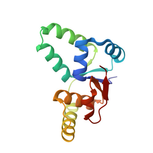
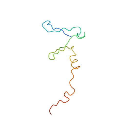
Crystal Structure of the VapBC Toxin-Antitoxin Complex from Shigella flexneri Reveals a Hetero-Octameric DNA-Binding Assembly.

Dienemann, C.Boggild, A.Winther, K.S.Gerdes, K.Brodersen, D.E.

(2011) J Mol Biology 414: 713-722

  • DOI: https://doi.org/10.1016/j.jmb.2011.10.024
  • Primary Citation Related Structures: 
    3TND

  • PubMed Abstract: 

    Toxin-antitoxin (TA) loci are common in archaea and prokaryotes and allow cells to rapidly adapt to changing environmental conditions through release of active regulators of metabolism. Many toxins are endonucleases that target cellular mRNA and tRNAs, while the antitoxins tightly wrap around the toxins to inhibit them under normal circumstances. The antitoxins also bind to operators in the promoter regions of the cognate TA operon and thereby regulate transcription. For enteric vapBC TA loci, the VapC toxins specifically cleave tRNA(fMet) and thus down-regulate protein synthesis. Here, we describe the crystal structure of the intact Shigella flexneri VapBC TA complex, determined to 2.7 Å resolution. Both in solution and in the crystal structure, four molecules of each protein combine to form a large and globular hetero-octameric assembly with SpoVT/AbrB-type DNA-binding domains at each end and a total molecular mass of about 100 kDa. The structure gives new insights into the inhibition of VapC toxins by VapB and provides the molecular basis for understanding transcriptional regulation through VapB dimerization.


  • Organizational Affiliation
    • Centre for mRNP Biogenesis and Metabolism, Department of Molecular Biology and Genetics, Aarhus University, Gustav Wieds Vej 10C, DK-8000 Aarhus C, Denmark.

Macromolecule Content 

  • Total Structure Weight: 98.06 kDa 
  • Atom Count: 6,542 
  • Modeled Residue Count: 797 
  • Deposited Residue Count: 852 
  • Unique protein chains: 2

Macromolecules

Find similar proteins by:|  3D Structure
Entity ID: 1
MoleculeChains  Sequence LengthOrganismDetailsImage
tRNA(fMet)-specific endonuclease VapC
A, C, E, G
132Shigella flexneriMutation(s): 0 
Gene Names: CP0245mvpAstborf2vapC
EC: 3.1
UniProt
Find proteins for O06662 (Shigella flexneri)
Explore O06662 
Go to UniProtKB:  O06662
Entity Groups
Sequence Clusters30% Identity50% Identity70% Identity90% Identity95% Identity100% Identity
UniProt GroupO06662
Sequence Annotations
Expand
Reference Sequence
Find similar proteins by:|  3D Structure
Entity ID: 2
MoleculeChains  Sequence LengthOrganismDetailsImage
Antitoxin VapB
B, D, F, H
81Shigella flexneriMutation(s): 0 
Gene Names: CP0246mvpTvapB
UniProt
Find proteins for O06663 (Shigella flexneri)
Explore O06663 
Go to UniProtKB:  O06663
Entity Groups
Sequence Clusters30% Identity50% Identity70% Identity90% Identity95% Identity100% Identity
UniProt GroupO06663
Sequence Annotations
Expand
Reference Sequence

Small Molecules

Ligands 2 Unique
IDChains Name / Formula / InChI Key2D Diagram3D Interactions
SO4

Query on SO4


Download:Ideal Coordinates CCD File
I [auth A]
K [auth B]
M [auth D]
N [auth D]
O [auth E]
I [auth A],
K [auth B],
M [auth D],
N [auth D],
O [auth E],
P [auth F],
Q [auth G],
R [auth H],
S [auth H],
T [auth H],
U [auth H]
SULFATE ION
O4 S
QAOWNCQODCNURD-UHFFFAOYSA-L
NA

Query on NA


Download:Ideal Coordinates CCD File
J [auth B],
L [auth D]
SODIUM ION
Na
FKNQFGJONOIPTF-UHFFFAOYSA-N

Experimental Data & Validation

Experimental Data

  • Method: X-RAY DIFFRACTION
  • Resolution: 2.70 Å
  • R-Value Free:  0.237 (Depositor), 0.231 (DCC) 
  • R-Value Work:  0.182 (Depositor), 0.177 (DCC) 
  • R-Value Observed: 0.185 (Depositor) 
Space Group: P 61 2 2
Unit Cell:
Length ( Å )Angle ( ˚ )
a = 91.403α = 90
b = 91.403β = 90
c = 549.09γ = 120
Software Package:
Software NamePurpose
DNAdata collection
SOLVEphasing
PHENIXrefinement
XDSdata reduction
XSCALEdata scaling

Structure Validation

View Full Validation Report


Entry History 

Deposition Data

Revision History  (Full details and data files)

  • Version 1.0: 2011-11-02
    Type: Initial release
  • Version 1.1: 2011-11-16
    Changes: Database references
  • Version 1.2: 2011-12-21
    Changes: Database references
  • Version 1.3: 2018-03-07
    Changes: Data collection
  • Version 1.4: 2024-02-28
    Changes: Data collection, Database references, Derived calculations