3TMN

THE BINDING OF L-VALYL-L-TRYPTOPHAN TO CRYSTALLINE THERMOLYSIN ILLUSTRATES THE MODE OF INTERACTION OF A PRODUCT OF PEPTIDE HYDROLYSIS


Experimental Data Snapshot

  • Method: X-RAY DIFFRACTION
  • Resolution: 1.70 Å
  • R-Value Observed: 0.173 

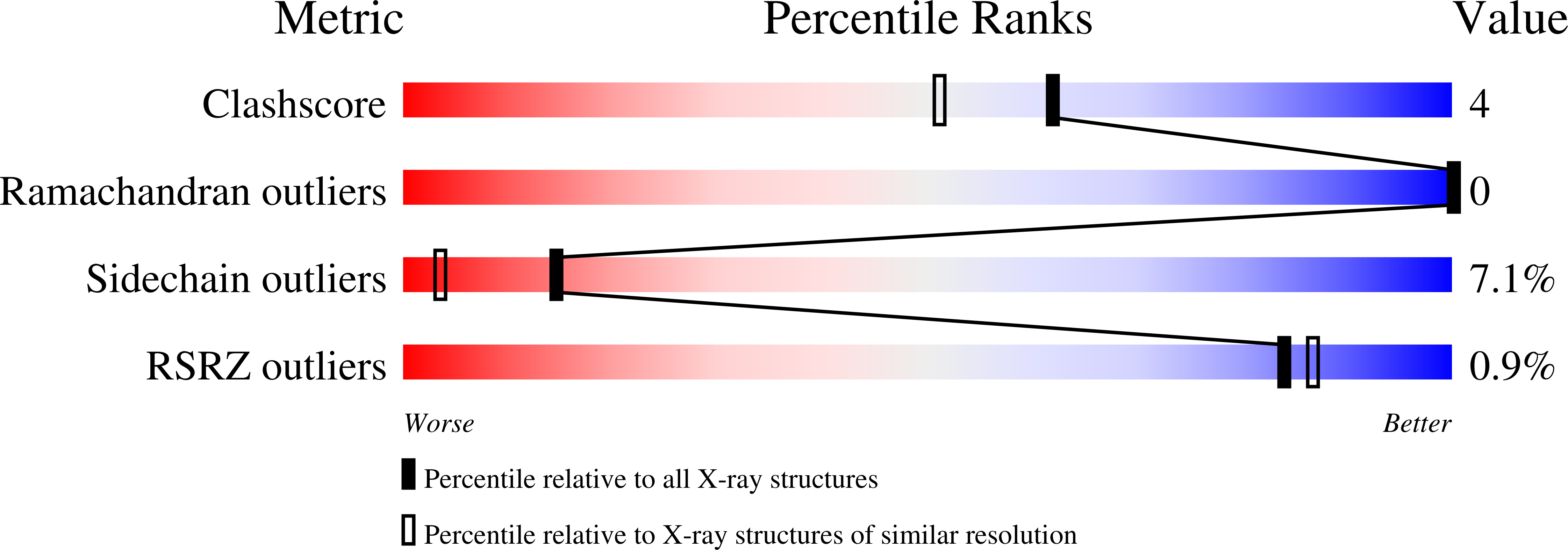
wwPDB Validation   3D Report Full Report


Ligand Structure Quality Assessment 


This is version 2.1 of the entry. See complete history


Literature

The binding of L-valyl-L-tryptophan to crystalline thermolysin illustrates the mode of interaction of a product of peptide hydrolysis.

Holden, H.M.Matthews, B.W.

(1988) J Biol Chem 263: 3256-3260

  • DOI: https://doi.org/10.2210/pdb3tmn/pdb
  • Primary Citation of Related Structures:  
    3TMN

  • PubMed Abstract: 

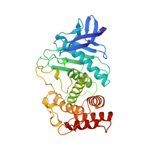
    Crystallographic analysis of the binding of mercaptoacetyl-L-valyl-L-tryptophan to thermolysin suggests that this inhibitor is hydrolyzed by the crystalline enzyme. The apparent product of hydrolysis, L-valyl-L-tryptophan (Val-Trp), occupies the S1'-S2' subsites of the active site, not the S1-S1' subsites as observed previously for the dipeptide L-alanyl-L-phenylalanine (Ala-Phe). The difference in binding of Val-Trp and Ala-Phe is consistent with the specificity requirements and preferences of thermolysin. The binding of Val-Trp illustrates the mode of interaction of one of the products of peptide hydrolysis. High resolution crystallographic refinement indicates that the valyl amino group makes three hydrogen bonds to the enzyme and to solvent and, in addition, is 2.8 A from the carboxylate of Glu-143. This is the first instance in which a direct interaction has been observed between Glu-143 and the scissile nitrogen. As such, the study directly supports the mechanism of action for thermolysin proposed by Hangauer et al. (Hangauer, D. G., Monzingo, A. F., and Matthews, B. W. (1984) Biochemistry 23, 5730-5741) and, by analogy, indirectly supports the similar mechanism proposed for carboxypeptidase A (Monzingo, A. F., and Matthews, B. W. (1984) Biochemistry 23, 5724-5729).


  • Organizational Affiliation

    Institute of Molecular Biology, University of Oregon, Eugene 97403.


Macromolecules
Find similar proteins by:  (by identity cutoff)  |  3D Structure
Entity ID: 1
MoleculeChains Sequence LengthOrganismDetailsImage
THERMOLYSINA [auth E]316Bacillus thermoproteolyticusMutation(s): 0 
EC: 3.4.24.27
UniProt
Find proteins for P00800 (Bacillus thermoproteolyticus)
Explore P00800 
Go to UniProtKB:  P00800
Entity Groups  
Sequence Clusters30% Identity50% Identity70% Identity90% Identity95% Identity100% Identity
UniProt GroupP00800
Sequence Annotations
Expand
  • Reference Sequence
Experimental Data & Validation

Experimental Data

  • Method: X-RAY DIFFRACTION
  • Resolution: 1.70 Å
  • R-Value Observed: 0.173 
  • Space Group: P 61 2 2
Unit Cell:
Length ( Å )Angle ( ˚ )
a = 94.1α = 90
b = 94.1β = 90
c = 131.4γ = 120
Software Package:
Software NamePurpose
TNTrefinement
OSCTSTdata reduction
AGROVATA/ROTAVATEdata scaling
FRODOmodel building

Structure Validation

View Full Validation Report


Ligand Structure Quality Assessment 


Entry History 

Deposition Data

Revision History  (Full details and data files)

  • Version 1.0: 1989-01-09
    Type: Initial release
  • Version 1.1: 2008-03-03
    Changes: Version format compliance
  • Version 1.2: 2011-07-13
    Changes: Advisory, Atomic model, Database references, Derived calculations, Non-polymer description, Structure summary, Version format compliance
  • Version 2.0: 2022-11-23
    Type: Remediation
    Changes: Advisory, Atomic model, Data collection, Database references, Derived calculations, Other
  • Version 2.1: 2024-05-22
    Changes: Data collection