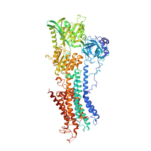
3TLM

Crystal Structure of Endoplasmic Reticulum Ca2+-ATPase (SERCA) From Bovine Muscle


Experimental Data Snapshot

  • Method: X-RAY DIFFRACTION
  • Resolution: 2.95 Å
  • R-Value Free: 0.277 
  • R-Value Work: 0.205 
  • R-Value Observed: 0.205 

Starting Model: experimental
View more details

wwPDB Validation   3D Report Full Report


Ligand Structure Quality Assessment 


This is version 1.2 of the entry. See complete history


Literature

Crystal structure of sarcoplasmic reticulum Ca(2+)-ATPase (SERCA) from bovine muscle.

Sacchetto, R.Bertipaglia, I.Giannetti, S.Cendron, L.Mascarello, F.Damiani, E.Carafoli, E.Zanotti, G.

(2012) J Struct Biol 178: 38-44

  • DOI: https://doi.org/10.1016/j.jsb.2012.02.008
  • Primary Citation of Related Structures:  
    3TLM

  • PubMed Abstract: 

    The SERCA pump, a membrane protein of about 110kDa, transports two Ca(2+) ions per ATP hydrolyzed from the cytoplasm to the lumen of the sarcoplasmic reticulum. In muscle cells, its ability to remove Ca(2+) from the cytosol induces relaxation. The transport mechanism employed by the enzyme from rabbit muscle has been extensively studied, and several crystal structures representing different conformational states are available. However, no structure of the pump from other sources is known. In this paper we describe the crystal structure of the bovine enzyme, crystallized in the E1 conformation and determined at 2.9Å resolution. The overall molecular model is very similar to that of the rabbit enzyme, as expected by the high amino acid sequence identity. Nevertheless, the bovine enzyme has reduced catalytic activity with respect to the rabbit enzyme. Subtle structural modifications, in particular in the region of the long loop that protrudes into the SR lumen connecting transmembrane α-helices M7 and M8, may explain the difference.


  • Organizational Affiliation

    Department of Experimental Veterinary Sciences, Viale dell'Università 16, 35020 Legnaro, Padua, Italy.


Macromolecules
Find similar proteins by:  (by identity cutoff)  |  3D Structure
Entity ID: 1
MoleculeChains Sequence LengthOrganismDetailsImage
Sarcoplasmic/endoplasmic reticulum calcium ATPase 1992Bos taurusMutation(s): 0 
EC: 3.6.3.8 (PDB Primary Data), 7.2.2.10 (UniProt)
Membrane Entity: Yes 
UniProt
Find proteins for Q0VCY0 (Bos taurus)
Explore Q0VCY0 
Go to UniProtKB:  Q0VCY0
Entity Groups  
Sequence Clusters30% Identity50% Identity70% Identity90% Identity95% Identity100% Identity
UniProt GroupQ0VCY0
Sequence Annotations
Expand
  • Reference Sequence
Experimental Data & Validation

Experimental Data

  • Method: X-RAY DIFFRACTION
  • Resolution: 2.95 Å
  • R-Value Free: 0.277 
  • R-Value Work: 0.205 
  • R-Value Observed: 0.205 
  • Space Group: C 1 2 1
Unit Cell:
Length ( Å )Angle ( ˚ )
a = 156.58α = 90
b = 75.26β = 108.3
c = 151.8γ = 90
Software Package:
Software NamePurpose
ADSCdata collection
PHASERphasing
CNSrefinement
MOSFLMdata reduction
SCALAdata scaling

Structure Validation

View Full Validation Report


Ligand Structure Quality Assessment 


Entry History 

Deposition Data

Revision History  (Full details and data files)

  • Version 1.0: 2012-03-21
    Type: Initial release
  • Version 1.1: 2012-04-25
    Changes: Database references
  • Version 1.2: 2023-09-13
    Changes: Data collection, Database references, Derived calculations, Refinement description