3THD | pdb_00003thd

Crystal structure of human beta-galactosidase in complex with 1-deoxygalactonojirimycin


Experimental Data Snapshot

  • Method: X-RAY DIFFRACTION
  • Resolution: 1.79 Å
  • R-Value Free: 
    0.228 (Depositor), 0.229 (DCC) 
  • R-Value Work: 
    0.198 (Depositor), 0.200 (DCC) 
  • R-Value Observed: 
    0.199 (Depositor) 

Starting Model: experimental
View more details

wwPDB Validation 3D Report Full Report

Validation slider image for 3THD

Ligand Structure Quality Assessment 


This is version 1.4 of the entry. See complete history

Literature

Crystal structure of human beta-galactosidase: structural basis of Gm1 gangliosidosis and morquio B diseases

Ohto, U.Usui, K.Ochi, T.Yuki, K.Satow, Y.Shimizu, T.

(2012) J Biological Chem 287: 1801-1812

  • DOI: https://doi.org/10.1074/jbc.M111.293795
  • Primary Citation Related Structures: 
    3THC, 3THD

  • PubMed Abstract: 

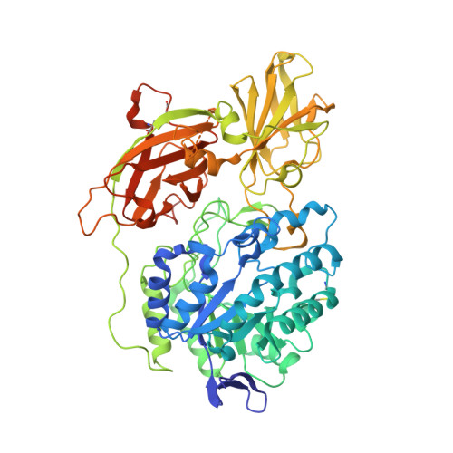
    G(M1) gangliosidosis and Morquio B are autosomal recessive lysosomal storage diseases associated with a neurodegenerative disorder or dwarfism and skeletal abnormalities, respectively. These diseases are caused by deficiencies in the lysosomal enzyme β-d-galactosidase (β-Gal), which lead to accumulations of the β-Gal substrates, G(M1) ganglioside, and keratan sulfate. β-Gal is an exoglycosidase that catalyzes the hydrolysis of terminal β-linked galactose residues. This study shows the crystal structures of human β-Gal in complex with its catalytic product galactose or with its inhibitor 1-deoxygalactonojirimycin. Human β-Gal is composed of a catalytic TIM barrel domain followed by β-domain 1 and β-domain 2. To gain structural insight into the molecular defects of β-Gal in the above diseases, the disease-causing mutations were mapped onto the three-dimensional structure. Finally, the possible causes of the diseases are discussed.


  • Organizational Affiliation
    • From the Graduate School of Pharmaceutical Sciences, University of Tokyo, 7-3-1 Hongo, Bunkyo-ku, Tokyo 113-0033, Japan.

Macromolecule Content 

  • Total Structure Weight: 300.26 kDa 
  • Atom Count: 21,969 
  • Modeled Residue Count: 2,414 
  • Deposited Residue Count: 2,616 
  • Unique protein chains: 1

Macromolecules

Find similar proteins by:|  3D Structure
Entity ID: 1
MoleculeChains  Sequence LengthOrganismDetailsImage
Beta-galactosidase
A, B, C, D
654Homo sapiensMutation(s): 0 
Gene Names: GLB1ELNR1
EC: 3.2.1.23
UniProt & NIH Common Fund Data Resources
Find proteins for P16278 (Homo sapiens)
Explore P16278 
Go to UniProtKB:  P16278
PHAROS:  P16278
GTEx:  ENSG00000170266 
Entity Groups
Sequence Clusters30% Identity50% Identity70% Identity90% Identity95% Identity100% Identity
UniProt GroupP16278
Glycosylation
Glycosylation Sites: 4Go to GlyGen: P16278-1
Sequence Annotations
Expand
Reference Sequence

Small Molecules

Ligands 5 Unique
IDChains Name / Formula / InChI Key2D Diagram3D Interactions
NAG

Query on NAG


Download:Ideal Coordinates CCD File
AA [auth C]
BA [auth C]
E [auth A]
F [auth A]
G [auth A]
AA [auth C],
BA [auth C],
E [auth A],
F [auth A],
G [auth A],
H [auth A],
IA [auth D],
JA [auth D],
KA [auth D],
LA [auth D],
O [auth B],
P [auth B],
Q [auth B],
R [auth B],
Y [auth C],
Z [auth C]
2-acetamido-2-deoxy-beta-D-glucopyranose
C8 H15 N O6
OVRNDRQMDRJTHS-FMDGEEDCSA-N
DGJ

Query on DGJ


Download:Ideal Coordinates CCD File
DA [auth C],
J [auth A],
NA [auth D],
T [auth B]
(2R,3S,4R,5S)-2-(hydroxymethyl)piperidine-3,4,5-triol
C6 H13 N O4
LXBIFEVIBLOUGU-DPYQTVNSSA-N
SO4

Query on SO4


Download:Ideal Coordinates CCD File
EA [auth C]
FA [auth C]
K [auth A]
L [auth A]
OA [auth D]
EA [auth C],
FA [auth C],
K [auth A],
L [auth A],
OA [auth D],
PA [auth D],
U [auth B],
V [auth B]
SULFATE ION
O4 S
QAOWNCQODCNURD-UHFFFAOYSA-L
EDO

Query on EDO


Download:Ideal Coordinates CCD File
GA [auth C]
HA [auth C]
M [auth A]
N [auth A]
QA [auth D]
GA [auth C],
HA [auth C],
M [auth A],
N [auth A],
QA [auth D],
RA [auth D],
W [auth B],
X [auth B]
1,2-ETHANEDIOL
C2 H6 O2
LYCAIKOWRPUZTN-UHFFFAOYSA-N
CL

Query on CL


Download:Ideal Coordinates CCD File
CA [auth C],
I [auth A],
MA [auth D],
S [auth B]
CHLORIDE ION
Cl
VEXZGXHMUGYJMC-UHFFFAOYSA-M
Binding Affinity Annotations 
IDSourceBinding Affinity
DGJ BindingDB:  3THD Ki: min: 4.47e+4, max: 6.18e+4 (nM) from 3 assay(s)
IC50: 9.00e+4 (nM) from 1 assay(s)

Experimental Data & Validation

Experimental Data

  • Method: X-RAY DIFFRACTION
  • Resolution: 1.79 Å
  • R-Value Free:  0.228 (Depositor), 0.229 (DCC) 
  • R-Value Work:  0.198 (Depositor), 0.200 (DCC) 
  • R-Value Observed: 0.199 (Depositor) 
Space Group: P 1 21 1
Unit Cell:
Length ( Å )Angle ( ˚ )
a = 94.792α = 90
b = 115.996β = 92.22
c = 140.327γ = 90
Software Package:
Software NamePurpose
MOLREPphasing
REFMACrefinement
HKL-2000data reduction
HKL-2000data scaling

Structure Validation

View Full Validation Report


Ligand Structure Quality Assessment 


Entry History 

Deposition Data

Revision History  (Full details and data files)

  • Version 1.0: 2011-12-07
    Type: Initial release
  • Version 1.1: 2013-07-10
    Changes: Database references
  • Version 1.2: 2020-07-29
    Type: Remediation
    Reason: Carbohydrate remediation
    Changes: Data collection, Derived calculations, Structure summary
  • Version 1.3: 2023-11-01
    Changes: Data collection, Database references, Refinement description, Structure summary
  • Version 1.4: 2024-11-13
    Changes: Structure summary