3TFL | pdb_00003tfl

LytR-Cps2a-Psr family protein with bound octaprenyl pyrophosphate lipid


Experimental Data Snapshot

  • Method: X-RAY DIFFRACTION
  • Resolution: 2.05 Å
  • R-Value Free: 
    0.242 (Depositor), 0.229 (DCC) 
  • R-Value Work: 
    0.191 (Depositor) 
  • R-Value Observed: 
    0.193 (Depositor) 

wwPDB Validation 3D Report Full Report

Validation slider image for 3TFL

Ligand Structure Quality Assessment 


This is version 1.2 of the entry. See complete history

Literature

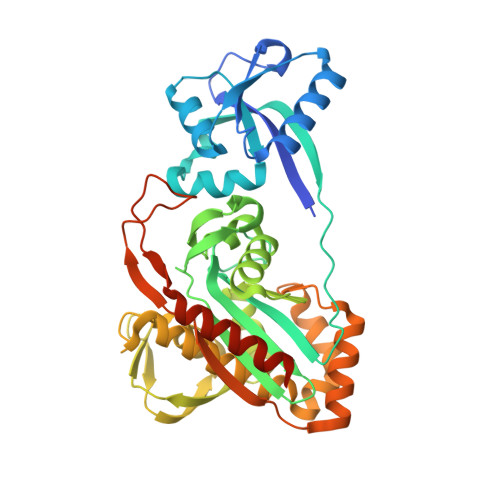
A widespread family of bacterial cell wall assembly proteins.

Kawai, Y.Marles-Wright, J.Cleverley, R.M.Emmins, R.Ishikawa, S.Kuwano, M.Heinz, N.Bui, N.K.Hoyland, C.N.Ogasawara, N.Lewis, R.J.Vollmer, W.Daniel, R.A.Errington, J.

(2011) EMBO J 30: 4931-4941

  • DOI: https://doi.org/10.1038/emboj.2011.358
  • Primary Citation Related Structures: 
    2XXP, 2XXQ, 3TEL, 3TEP, 3TFL

  • PubMed Abstract: 

    Teichoic acids and acidic capsular polysaccharides are major anionic cell wall polymers (APs) in many bacteria, with various critical cell functions, including maintenance of cell shape and structural integrity, charge and cation homeostasis, and multiple aspects of pathogenesis. We have identified the widespread LytR-Cps2A-Psr (LCP) protein family, of previously unknown function, as novel enzymes required for AP synthesis. Structural and biochemical analysis of several LCP proteins suggest that they carry out the final step of transferring APs from their lipid-linked precursor to cell wall peptidoglycan (PG). In Bacillus subtilis, LCP proteins are found in association with the MreB cytoskeleton, suggesting that MreB proteins coordinate the insertion of the major polymers, PG and AP, into the cell wall.


  • Organizational Affiliation
    • Centre for Bacterial Cell Biology, Medical School, Newcastle University, Newcastle upon Tyne, UK.

Macromolecule Content 

  • Total Structure Weight: 45.09 kDa 
  • Atom Count: 3,107 
  • Modeled Residue Count: 371 
  • Deposited Residue Count: 398 
  • Unique protein chains: 1

Macromolecules

Find similar proteins by:|  3D Structure
Entity ID: 1
MoleculeChains  Sequence LengthOrganismDetailsImage
CPS2A, Integral membrane regulatory protein Wzg398Streptococcus pneumoniaeMutation(s): 0 
Gene Names: SPC02_0004wzg
UniProt
Find proteins for Q4K376 (Streptococcus pneumoniae)
Explore Q4K376 
Go to UniProtKB:  Q4K376
Entity Groups
Sequence Clusters30% Identity50% Identity70% Identity90% Identity95% Identity100% Identity
UniProt GroupQ4K376
Sequence Annotations
Expand
Reference Sequence

Small Molecules

Ligands 2 Unique
IDChains Name / Formula / InChI Key2D Diagram3D Interactions
ZTP

Query on ZTP


Download:Ideal Coordinates CCD File
C [auth A](2Z,6Z,10Z,14Z,18Z,22Z,26Z)-3,7,11,15,19,23,27,31-octamethyldotriaconta-2,6,10,14,18,22,26,30-octaen-1-yl trihydrogen diphosphate
C40 H68 O7 P2
IKKLDISSULFFQO-DLTKSLTJSA-N
EDO

Query on EDO


Download:Ideal Coordinates CCD File
B [auth A]
D [auth A]
E [auth A]
F [auth A]
G [auth A]
B [auth A],
D [auth A],
E [auth A],
F [auth A],
G [auth A],
H [auth A]
1,2-ETHANEDIOL
C2 H6 O2
LYCAIKOWRPUZTN-UHFFFAOYSA-N

Experimental Data & Validation

Experimental Data

  • Method: X-RAY DIFFRACTION
  • Resolution: 2.05 Å
  • R-Value Free:  0.242 (Depositor), 0.229 (DCC) 
  • R-Value Work:  0.191 (Depositor) 
  • R-Value Observed: 0.193 (Depositor) 
Space Group: P 43 21 2
Unit Cell:
Length ( Å )Angle ( ˚ )
a = 73.348α = 90
b = 73.348β = 90
c = 163.869γ = 90
Software Package:
Software NamePurpose
GDAdata collection
PHASERphasing
REFMACrefinement
XDSdata reduction
SCALAdata scaling

Structure Validation

View Full Validation Report


Ligand Structure Quality Assessment 


Entry History 

Revision History  (Full details and data files)

  • Version 1.0: 2011-10-12
    Type: Initial release
  • Version 1.1: 2013-01-09
    Changes: Database references
  • Version 1.2: 2024-02-28
    Changes: Data collection, Database references, Derived calculations