3TFK | pdb_00003tfk

42F3-p4B10/H2-Ld


Experimental Data Snapshot

  • Method: X-RAY DIFFRACTION
  • Resolution: 2.75 Å
  • R-Value Free: 
    0.291 (Depositor), 0.281 (DCC) 
  • R-Value Work: 
    0.247 (Depositor), 0.240 (DCC) 
  • R-Value Observed: 
    0.250 (Depositor) 

Starting Model: experimental
View more details

wwPDB Validation 3D Report Full Report

Validation slider image for 3TFK

This is version 1.3 of the entry. See complete history

Literature

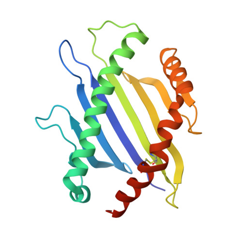
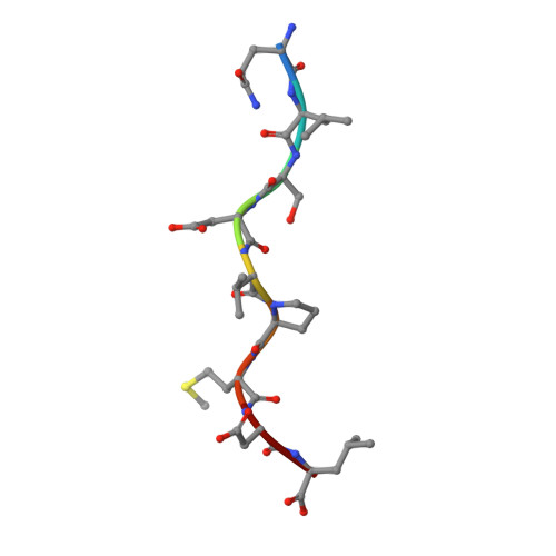
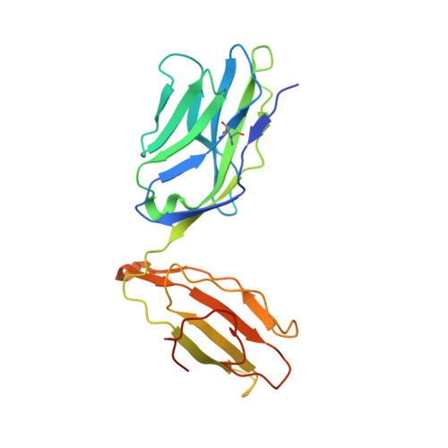
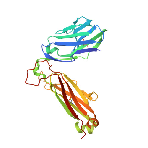
T cell receptor signaling is limited by docking geometry to peptide-major histocompatibility complex.

Adams, J.J.Narayanan, S.Liu, B.Birnbaum, M.E.Kruse, A.C.Bowerman, N.A.Chen, W.Levin, A.M.Connolly, J.M.Zhu, C.Kranz, D.M.Garcia, K.C.

(2011) Immunity 35: 681-693

  • DOI: https://doi.org/10.1016/j.immuni.2011.09.013
  • Primary Citation Related Structures: 
    3TF7, 3TFK, 3TJH, 3TPU

  • PubMed Abstract: 

    T cell receptor (TCR) engagement of peptide-major histocompatibility complex (pMHC) is essential to adaptive immunity, but it is unknown whether TCR signaling responses are influenced by the binding topology of the TCR-peptide-MHC complex. We developed yeast-displayed pMHC libraries that enabled us to identify new peptide sequences reactive with a single TCR. Structural analysis showed that four peptides bound to the TCR with distinct 3D and 2D affinities using entirely different binding chemistries. Three of the peptides that shared a common docking mode, where key TCR-MHC germline interactions are preserved, induced TCR signaling. The fourth peptide failed to induce signaling and was recognized in a substantially different TCR-MHC binding mode that apparently exceeded geometric tolerances compatible with signaling. We suggest that the stereotypical TCR-MHC docking paradigm evolved from productive signaling geometries and that TCR signaling can be modulated by peptides that are recognized in alternative TCR-pMHC binding orientations.


  • Organizational Affiliation
    • Howard Hughes Medical Institute, and Department of Molecular and Cellular Physiology, Stanford University School of Medicine, Stanford, CA 94305, USA.

Macromolecule Content 

  • Total Structure Weight: 72.78 kDa 
  • Atom Count: 4,982 
  • Modeled Residue Count: 623 
  • Deposited Residue Count: 644 
  • Unique protein chains: 4

Macromolecules

Find similar proteins by:|  3D Structure
Entity ID: 1
MoleculeChains  Sequence LengthOrganismDetailsImage
H2-Ld SBM2180Mus musculusMutation(s): 0 
UniProt
Find proteins for P01897 (Mus musculus)
Explore P01897 
Go to UniProtKB:  P01897
Entity Groups
Sequence Clusters30% Identity50% Identity70% Identity90% Identity95% Identity100% Identity
UniProt GroupP01897
Sequence Annotations
Expand
Reference Sequence
Find similar proteins by:  Sequence   |   3D Structure  
Entity ID: 2
MoleculeChains  Sequence LengthOrganismDetailsImage
p4B10 peptide9N/AMutation(s): 0 
Find similar proteins by:|  3D Structure
Entity ID: 3
MoleculeChains  Sequence LengthOrganismDetailsImage
42F3 alpha212Mus musculusHomo sapiens
This entity is chimeric
Mutation(s): 0 
Find similar proteins by:|  3D Structure
Entity ID: 4
MoleculeChains  Sequence LengthOrganismDetailsImage
42F3 beta243Mus musculusHomo sapiens
This entity is chimeric
Mutation(s): 0 

Small Molecules

Ligands 1 Unique
IDChains Name / Formula / InChI Key2D Diagram3D Interactions
NO3

Query on NO3


Download:Ideal Coordinates CCD File
E [auth D]NITRATE ION
N O3
NHNBFGGVMKEFGY-UHFFFAOYSA-N

Experimental Data & Validation

Experimental Data

  • Method: X-RAY DIFFRACTION
  • Resolution: 2.75 Å
  • R-Value Free:  0.291 (Depositor), 0.281 (DCC) 
  • R-Value Work:  0.247 (Depositor), 0.240 (DCC) 
  • R-Value Observed: 0.250 (Depositor) 
Space Group: P 61 2 2
Unit Cell:
Length ( Å )Angle ( ˚ )
a = 102.09α = 90
b = 102.09β = 90
c = 323.081γ = 120
Software Package:
Software NamePurpose
Blu-Icedata collection
PHASERphasing
PHENIXrefinement
MOSFLMdata reduction
SCALAdata scaling

Structure Validation

View Full Validation Report


Entry History 

Deposition Data

Revision History  (Full details and data files)

  • Version 1.0: 2011-12-07
    Type: Initial release
  • Version 1.1: 2012-01-18
    Changes: Database references
  • Version 1.2: 2023-09-13
    Changes: Data collection, Database references, Derived calculations, Refinement description
  • Version 1.3: 2024-10-16
    Changes: Structure summary