3TEJ | pdb_00003tej

Crystal structure of a domain fragment involved in peptide natural product biosynthesis


Experimental Data Snapshot

  • Method: X-RAY DIFFRACTION
  • Resolution: 1.90 Å
  • R-Value Free: 
    0.227 (Depositor), 0.227 (DCC) 
  • R-Value Work: 
    0.206 (Depositor), 0.211 (DCC) 
  • R-Value Observed: 
    0.206 (Depositor) 

wwPDB Validation 3D Report Full Report

Validation slider image for 3TEJ

This is version 1.1 of the entry. See complete history

Literature

Structural basis for phosphopantetheinyl carrier domain interactions in the terminal module of nonribosomal peptide synthetases.

Liu, Y.Zheng, T.Bruner, S.D.

(2011) Chem Biol 18: 1482-1488

  • DOI: https://doi.org/10.1016/j.chembiol.2011.09.018
  • Primary Citation Related Structures: 
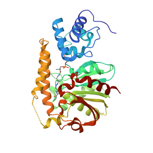
    3TEJ

  • PubMed Abstract: 

    Phosphopantetheine-modified carrier domains play a central role in the template-directed, biosynthesis of several classes of primary and secondary metabolites. Fatty acids, polyketides, and nonribosomal peptides are constructed on multidomain enzyme assemblies using phosphopantetheinyl thioester-linked carrier domains to traffic and activate building blocks. The carrier domain is a dynamic component of the process, shuttling pathway intermediates to sequential enzyme active sites. Here, we report an approach to structurally fix carrier domain/enzyme constructs suitable for X-ray crystallographic analysis. The structure of a two-domain construct of Escherichia coli EntF was determined with a conjugated phosphopantetheinyl-based inhibitor. The didomain structure is locked in an active orientation relevant to the chemistry of nonribosomal peptide biosynthesis. This structure provides details into the interaction of phosphopantetheine arm with the carrier domain and the active site of the thioesterase domain.


  • Organizational Affiliation
    • Department of Chemistry, Boston College, Chestnut Hill, MA 02167, USA.

Macromolecule Content 

  • Total Structure Weight: 72.27 kDa 
  • Atom Count: 4,855 
  • Modeled Residue Count: 606 
  • Deposited Residue Count: 658 
  • Unique protein chains: 1

Macromolecules

Find similar proteins by:|  3D Structure
Entity ID: 1
MoleculeChains  Sequence LengthOrganismDetailsImage
Enterobactin synthase component F
A, B
329Escherichia coli K-12Mutation(s): 0 
Gene Names: entFb0586JW0578
EC: 2.7.7 (PDB Primary Data), 6.2.1.72 (UniProt), 6.3.2.14 (UniProt)
UniProt
Find proteins for P11454 (Escherichia coli (strain K12))
Explore P11454 
Go to UniProtKB:  P11454
Entity Groups
Sequence Clusters30% Identity50% Identity70% Identity90% Identity95% Identity100% Identity
UniProt GroupP11454
Sequence Annotations
Expand
Reference Sequence

Small Molecules

Modified Residues  1 Unique
IDChains TypeFormula2D DiagramParent
UF0
Query on UF0
A, B
L-PEPTIDE LINKINGC16 H31 N4 O11 PSER

Experimental Data & Validation

Experimental Data

  • Method: X-RAY DIFFRACTION
  • Resolution: 1.90 Å
  • R-Value Free:  0.227 (Depositor), 0.227 (DCC) 
  • R-Value Work:  0.206 (Depositor), 0.211 (DCC) 
  • R-Value Observed: 0.206 (Depositor) 
Space Group: P 21 21 21
Unit Cell:
Length ( Å )Angle ( ˚ )
a = 69.4α = 90
b = 90.36β = 90
c = 97.53γ = 90
Software Package:
Software NamePurpose
CNSrefinement
PDB_EXTRACTdata extraction
HKL-2000data reduction
HKL-2000data scaling
SOLVEphasing

Structure Validation

View Full Validation Report


Entry History 

Deposition Data

Revision History  (Full details and data files)

  • Version 1.0: 2012-01-18
    Type: Initial release
  • Version 1.1: 2025-03-26
    Changes: Data collection, Database references, Derived calculations, Structure summary