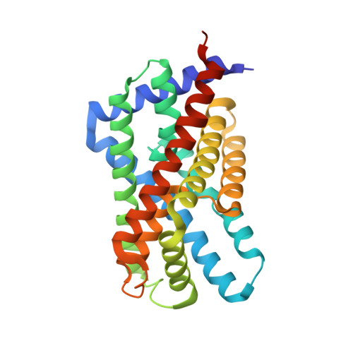
3TE2

Crystal structure of HSC K16S


Experimental Data Snapshot

  • Method: X-RAY DIFFRACTION
  • Resolution: 2.30 Å
  • R-Value Free: 0.230 
  • R-Value Work: 0.185 
  • R-Value Observed: 0.187 

Starting Model: experimental
View more details

wwPDB Validation   3D Report Full Report


Ligand Structure Quality Assessment 


This is version 1.4 of the entry. See complete history


Literature

Identification and characterization of a bacterial hydrosulphide ion channel.

Czyzewski, B.K.Wang, D.N.

(2012) Nature 483: 494-497

  • DOI: https://doi.org/10.1038/nature10881
  • Primary Citation of Related Structures:  
    3TDO, 3TDP, 3TDR, 3TDS, 3TDX, 3TE0, 3TE1, 3TE2

  • PubMed Abstract: 

    The hydrosulphide ion (HS(-)) and its undissociated form, hydrogen sulphide (H(2)S), which are believed to have been critical to the origin of life on Earth, remain important in physiology and cellular signalling. As a major metabolite in anaerobic bacterial growth, hydrogen sulphide is a product of both assimilatory and dissimilatory sulphate reduction. These pathways can reduce various oxidized sulphur compounds including sulphate, sulphite and thiosulphate. The dissimilatory sulphate reduction pathway uses this molecule as the terminal electron acceptor for anaerobic respiration, in which process it produces excess amounts of H(2)S (ref. 4). The reduction of sulphite is a key intermediate step in all sulphate reduction pathways. In Clostridium and Salmonella, an inducible sulphite reductase is directly linked to the regeneration of NAD(+), which has been suggested to have a role in energy production and growth, as well as in the detoxification of sulphite. Above a certain concentration threshold, both H(2)S and HS(-) inhibit cell growth by binding the metal centres of enzymes and cytochrome oxidase, necessitating a release mechanism for the export of this toxic metabolite from the cell. Here we report the identification of a hydrosulphide ion channel in the pathogen Clostridium difficile through a combination of genetic, biochemical and functional approaches. The HS(-) channel is a member of the formate/nitrite transport family, in which about 50 hydrosulphide ion channels form a third subfamily alongside those for formate (FocA) and for nitrite (NirC). The hydrosulphide ion channel is permeable to formate and nitrite as well as to HS(-) ions. Such polyspecificity can be explained by the conserved ion selectivity filter observed in the channel's crystal structure. The channel has a low open probability and is tightly regulated, to avoid decoupling of the membrane proton gradient.


  • Organizational Affiliation

    The Helen L. and Martin S. Kimmel Center for Biology and Medicine at the Skirball Institute of Biomolecular Medicine, New York University School of Medicine, 540 First Avenue, New York, New York 10016, USA.


Macromolecules
Find similar proteins by:  (by identity cutoff)  |  3D Structure
Entity ID: 1
MoleculeChains Sequence LengthOrganismDetailsImage
formate/nitrite transporter
A, B, C, D, E
268Clostridioides difficile 630Mutation(s): 1 
Gene Names: CD630_22300Putative formate/nitrite transporter
Membrane Entity: Yes 
UniProt
Find proteins for Q186B7 (Clostridioides difficile (strain 630))
Explore Q186B7 
Go to UniProtKB:  Q186B7
Entity Groups  
Sequence Clusters30% Identity50% Identity70% Identity90% Identity95% Identity100% Identity
UniProt GroupQ186B7
Sequence Annotations
Expand
  • Reference Sequence
Small Molecules
Ligands 2 Unique
IDChains Name / Formula / InChI Key2D Diagram3D Interactions
BOG
Query on BOG

Download Ideal Coordinates CCD File 
G [auth A]
H [auth A]
J [auth B]
K [auth B]
L [auth B]
G [auth A],
H [auth A],
J [auth B],
K [auth B],
L [auth B],
O [auth D],
Q [auth E]
octyl beta-D-glucopyranoside
C14 H28 O6
HEGSGKPQLMEBJL-RKQHYHRCSA-N
PG4
Query on PG4

Download Ideal Coordinates CCD File 
F [auth A],
I [auth B],
M [auth C],
N [auth D],
P [auth E]
TETRAETHYLENE GLYCOL
C8 H18 O5
UWHCKJMYHZGTIT-UHFFFAOYSA-N
Experimental Data & Validation

Experimental Data

  • Method: X-RAY DIFFRACTION
  • Resolution: 2.30 Å
  • R-Value Free: 0.230 
  • R-Value Work: 0.185 
  • R-Value Observed: 0.187 
  • Space Group: P 21 21 21
Unit Cell:
Length ( Å )Angle ( ˚ )
a = 98.689α = 90
b = 118.704β = 90
c = 150.672γ = 90
Software Package:
Software NamePurpose
DENZOdata reduction
SCALEPACKdata scaling
PHASERphasing
PHENIXrefinement
PDB_EXTRACTdata extraction

Structure Validation

View Full Validation Report


Ligand Structure Quality Assessment 


Entry History 

Deposition Data

Revision History  (Full details and data files)

  • Version 1.0: 2012-03-07
    Type: Initial release
  • Version 1.1: 2012-04-04
    Changes: Database references
  • Version 1.2: 2018-01-24
    Changes: Structure summary
  • Version 1.3: 2020-07-29
    Type: Remediation
    Reason: Carbohydrate remediation
    Changes: Data collection, Database references, Derived calculations, Structure summary
  • Version 1.4: 2023-09-13
    Changes: Data collection, Database references, Refinement description, Structure summary