3TCQ | pdb_00003tcq

Crystal Structure of matrix protein VP40 from Ebola virus Sudan


Experimental Data Snapshot

  • Method: X-RAY DIFFRACTION
  • Resolution: 1.60 Å
  • R-Value Free: 
    0.221 (Depositor), 0.238 (DCC) 
  • R-Value Work: 
    0.194 (Depositor), 0.213 (DCC) 
  • R-Value Observed: 
    0.196 (Depositor) 

Starting Model: experimental
View more details

wwPDB Validation 3D Report Full Report

Validation slider image for 3TCQ

This is version 1.4 of the entry. See complete history

Literature

High-resolution Crystal Structure of Dimeric VP40 From Sudan ebolavirus.

Clifton, M.C.Bruhn, J.F.Atkins, K.Webb, T.L.Baydo, R.O.Raymond, A.Lorimer, D.D.Edwards, T.E.Myler, P.J.Saphire, E.O.

(2015) J Infect Dis 212 Suppl 2: S167-S171

  • DOI: https://doi.org/10.1093/infdis/jiv090
  • Primary Citation Related Structures: 
    3TCQ

  • PubMed Abstract: 

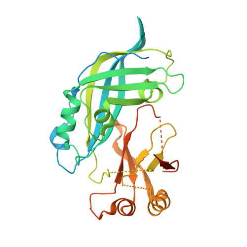
    Ebolaviruses cause severe hemorrhagic fever. Central to the Ebola life cycle is the matrix protein VP40, which oligomerizes and drives viral budding. Here we present the crystal structure of the Sudan virus (SUDV) matrix protein. This structure is higher resolution (1.6 Å) than previously achievable. Despite differences in the protein purification, we find that it still forms a stable dimer in solution, as was noted for other Ebola VP40s. Although the N-terminal domain interface by which VP40 dimerizes is conserved between Ebola virus and SUDV, the C-terminal domain interface by which VP40 dimers may further assemble is significantly smaller in this SUDV assembly.


  • Organizational Affiliation
    • Seattle Structural Genomics Center for Infectious Disease Beryllium, Bedford, Massachusetts.

Macromolecule Content 

  • Total Structure Weight: 35.51 kDa 
  • Atom Count: 1,970 
  • Modeled Residue Count: 239 
  • Deposited Residue Count: 326 
  • Unique protein chains: 1

Macromolecules

Find similar proteins by:|  3D Structure
Entity ID: 1
MoleculeChains  Sequence LengthOrganismDetailsImage
Matrix protein VP40326Sudan virus - Boniface, Sudan,1976Mutation(s): 0 
Gene Names: VP40SEBOVgp3
UniProt
Find proteins for B0LPL6 (Sudan ebolavirus)
Explore B0LPL6 
Go to UniProtKB:  B0LPL6
Entity Groups
Sequence Clusters30% Identity50% Identity70% Identity90% Identity95% Identity100% Identity
UniProt GroupB0LPL6
Sequence Annotations
Expand
Reference Sequence

Experimental Data & Validation

Experimental Data

  • Method: X-RAY DIFFRACTION
  • Resolution: 1.60 Å
  • R-Value Free:  0.221 (Depositor), 0.238 (DCC) 
  • R-Value Work:  0.194 (Depositor), 0.213 (DCC) 
  • R-Value Observed: 0.196 (Depositor) 
Space Group: C 1 2 1
Unit Cell:
Length ( Å )Angle ( ˚ )
a = 63.982α = 90
b = 90.94β = 95.86
c = 48.684γ = 90
Software Package:
Software NamePurpose
DENZOdata reduction
SCALEPACKdata scaling
PHASERphasing
REFMACrefinement
PDB_EXTRACTdata extraction
HKL-2000data reduction
HKL-2000data scaling

Structure Validation

View Full Validation Report


Entry History 

Revision History  (Full details and data files)

  • Version 1.0: 2012-10-03
    Type: Initial release
  • Version 1.1: 2016-01-20
    Changes: Database references
  • Version 1.2: 2017-11-08
    Changes: Refinement description
  • Version 1.3: 2023-09-13
    Changes: Data collection, Database references, Refinement description
  • Version 1.4: 2025-10-22
    Changes: Structure summary