3TCN | pdb_00003tcn

Crystal structures of Peptidyl-tRNA hydrolase from Mycobacterium tuberculosis - Form 2 grown in presence of Pentaglycine


Experimental Data Snapshot

  • Method: X-RAY DIFFRACTION
  • Resolution: 2.30 Å
  • R-Value Free: 
    0.228 (Depositor), 0.228 (DCC) 
  • R-Value Work: 
    0.180 (Depositor), 0.180 (DCC) 

wwPDB Validation 3D Report Full Report

Validation slider image for 3TCN

This is version 1.1 of the entry. See complete history

Literature

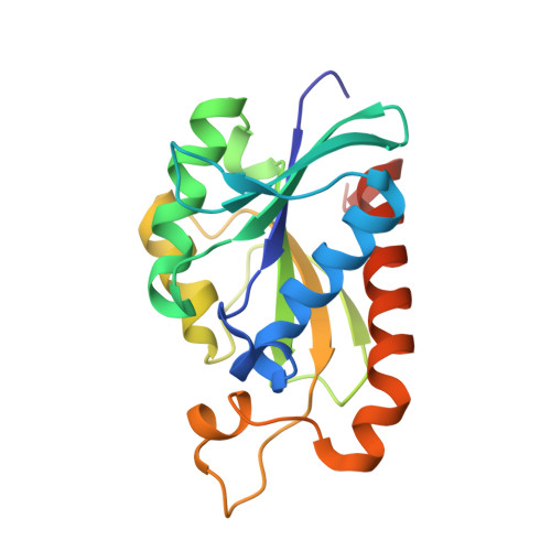
Structures of new crystal forms of Mycobacterium tuberculosis peptidyl-tRNA hydrolase and functionally important plasticity of the molecule

Selvaraj, M.Ahmad, R.Varshney, U.Vijayan, M.

(2012) Acta Crystallogr Sect F Struct Biol Cryst Commun 68: 124-128

  • DOI: https://doi.org/10.1107/S1744309111052341
  • Primary Citation Related Structures: 
    3TCK, 3TCN, 3TD2, 3TD6

  • PubMed Abstract: 

    The X-ray structures of new crystal forms of peptidyl-tRNA hydrolase from M. tuberculosis reported here and the results of previous X-ray studies of the enzyme from different sources provide a picture of the functionally relevant plasticity of the protein molecule. The new X-ray results confirm the connection deduced previously between the closure of the lid at the peptide-binding site and the opening of the gate that separates the peptide-binding and tRNA-binding sites. The plasticity of the molecule indicated by X-ray structures is in general agreement with that deduced from the available solution NMR results. The correlation between the lid and the gate movements is not, however, observed in the NMR structure.


  • Organizational Affiliation
    • Molecular Biophysics Unit, Indian Institute of Science, Bangalore, India.

Macromolecule Content 

  • Total Structure Weight: 40.97 kDa 
  • Atom Count: 3,114 
  • Modeled Residue Count: 371 
  • Deposited Residue Count: 382 
  • Unique protein chains: 1

Macromolecules

Find similar proteins by:|  3D Structure
Entity ID: 1
MoleculeChains  Sequence LengthOrganismDetailsImage
Peptidyl-tRNA hydrolase
A, B
191Mycobacterium tuberculosis H37RvMutation(s): 0 
Gene Names: MT1042MTCY10G2.35pthRv1014c
EC: 3.1.1.29
UniProt
Find proteins for P9WHN7 (Mycobacterium tuberculosis (strain ATCC 25618 / H37Rv))
Explore P9WHN7 
Go to UniProtKB:  P9WHN7
Entity Groups
Sequence Clusters30% Identity50% Identity70% Identity90% Identity95% Identity100% Identity
UniProt GroupP9WHN7
Sequence Annotations
Expand
Reference Sequence

Experimental Data & Validation

Experimental Data

  • Method: X-RAY DIFFRACTION
  • Resolution: 2.30 Å
  • R-Value Free:  0.228 (Depositor), 0.228 (DCC) 
  • R-Value Work:  0.180 (Depositor), 0.180 (DCC) 
Space Group: P 1 21 1
Unit Cell:
Length ( Å )Angle ( ˚ )
a = 35.74α = 90
b = 73.47β = 93.48
c = 59.95γ = 90
Software Package:
Software NamePurpose
MAR345dtbdata collection
PHASERphasing
CNSrefinement
MOSFLMdata reduction
SCALAdata scaling

Structure Validation

View Full Validation Report


Entry History 

Deposition Data

Revision History  (Full details and data files)

  • Version 1.0: 2012-02-15
    Type: Initial release
  • Version 1.1: 2024-03-20
    Changes: Data collection, Database references