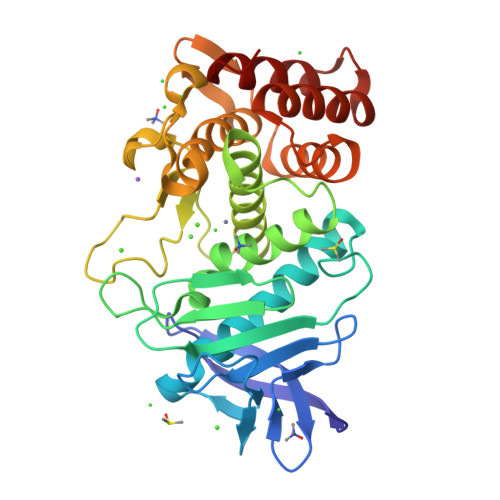
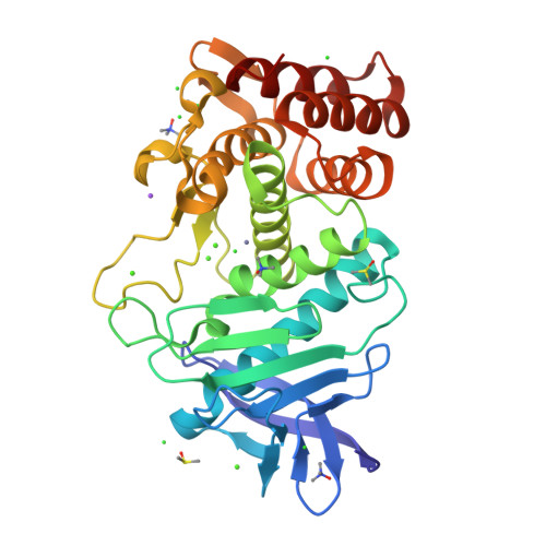
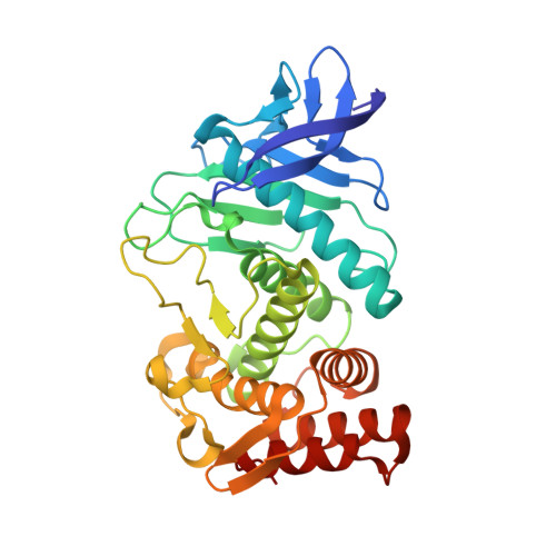
3T2H

Tetragonal thermolysin in the presence of TMAO


Experimental Data Snapshot

  • Method: X-RAY DIFFRACTION
  • Resolution: 1.95 Å
  • R-Value Free: 0.183 
  • R-Value Work: 0.154 
  • R-Value Observed: 0.156 

Starting Model: experimental
View more details

wwPDB Validation   3D Report Full Report


This is version 1.3 of the entry. See complete history


Literature

The use of trimethylamine N-oxide as a primary precipitating agent and related methylamine osmolytes as cryoprotective agents for macromolecular crystallography.

Marshall, H.Venkat, M.Hti Lar Seng, N.S.Cahn, J.Juers, D.H.

(2012) Acta Crystallogr D Biol Crystallogr 68: 69-81

  • DOI: https://doi.org/10.1107/S0907444911050360

  • PubMed Abstract: 

    Both crystallization and cryoprotection are often bottlenecks for high-resolution X-ray structure determination of macromolecules. Methylamine osmolytes are known stabilizers of protein structure. One such osmolyte, trimethylamine N-oxide (TMAO), has seen occasional use as an additive to improve macromolecular crystal quality and has recently been shown to be an effective cryoprotective agent for low-temperature data collection. Here, TMAO and the related osmolytes sarcosine and betaine are investigated as primary precipitating agents for protein crystal growth. Crystallization experiments were undertaken with 14 proteins. Using TMAO, seven proteins crystallized in a total of 13 crystal forms, including a new tetragonal crystal form of trypsin. The crystals diffracted well, and eight of the 13 crystal forms could be effectively cryocooled as grown with TMAO as an in situ cryoprotective agent. Sarcosine and betaine produced crystals of four and two of the 14 proteins, respectively. In addition to TMAO, sarcosine and betaine were effective post-crystallization cryoprotective agents for two different crystal forms of thermolysin. Precipitation reactions of TMAO with several transition-metal ions (Fe(3+), Co(2+), Cu(2+) and Zn(2+)) did not occur with sarcosine or betaine and were inhibited for TMAO at lower pH. Structures of proteins from TMAO-grown crystals and from crystals soaked in TMAO, sarcosine or betaine were determined, showing osmolyte binding in five of the 12 crystals tested. When an osmolyte was shown to bind, it did so near the protein surface, interacting with water molecules, side chains and backbone atoms, often at crystal contacts.


  • Organizational Affiliation

    Program in Biochemistry, Biophysics and Molecular Biology, Whitman College, Walla Walla, Washington, USA.


Macromolecules
Find similar proteins by:  (by identity cutoff)  |  3D Structure
Entity ID: 1
MoleculeChains Sequence LengthOrganismDetailsImage
ThermolysinA [auth E]316Bacillus thermoproteolyticusMutation(s): 0 
EC: 3.4.24.27
UniProt
Find proteins for P00800 (Bacillus thermoproteolyticus)
Explore P00800 
Go to UniProtKB:  P00800
Entity Groups  
Sequence Clusters30% Identity50% Identity70% Identity90% Identity95% Identity100% Identity
UniProt GroupP00800
Sequence Annotations
Expand
  • Reference Sequence
Small Molecules
Ligands 6 Unique
IDChains Name / Formula / InChI Key2D Diagram3D Interactions
DMS
Query on DMS

Download Ideal Coordinates CCD File 
T [auth E],
U [auth E]
DIMETHYL SULFOXIDE
C2 H6 O S
IAZDPXIOMUYVGZ-UHFFFAOYSA-N
TMO
Query on TMO

Download Ideal Coordinates CCD File 
Q [auth E],
R [auth E],
S [auth E]
trimethylamine oxide
C3 H9 N O
UYPYRKYUKCHHIB-UHFFFAOYSA-N
ZN
Query on ZN

Download Ideal Coordinates CCD File 
F [auth E],
G [auth E]
ZINC ION
Zn
PTFCDOFLOPIGGS-UHFFFAOYSA-N
CA
Query on CA

Download Ideal Coordinates CCD File 
B [auth E],
C [auth E],
D [auth E],
E
CALCIUM ION
Ca
BHPQYMZQTOCNFJ-UHFFFAOYSA-N
CL
Query on CL

Download Ideal Coordinates CCD File 
H [auth E]
I [auth E]
J [auth E]
K [auth E]
L [auth E]
H [auth E],
I [auth E],
J [auth E],
K [auth E],
L [auth E],
M [auth E],
N [auth E],
O [auth E]
CHLORIDE ION
Cl
VEXZGXHMUGYJMC-UHFFFAOYSA-M
NA
Query on NA

Download Ideal Coordinates CCD File 
P [auth E]SODIUM ION
Na
FKNQFGJONOIPTF-UHFFFAOYSA-N
Experimental Data & Validation

Experimental Data

  • Method: X-RAY DIFFRACTION
  • Resolution: 1.95 Å
  • R-Value Free: 0.183 
  • R-Value Work: 0.154 
  • R-Value Observed: 0.156 
  • Space Group: P 41 21 2
Unit Cell:
Length ( Å )Angle ( ˚ )
a = 96.892α = 90
b = 96.892β = 90
c = 105.549γ = 90
Software Package:
Software NamePurpose
SCALAdata scaling
REFMACrefinement
PDB_EXTRACTdata extraction
CrysalisProdata reduction

Structure Validation

View Full Validation Report


Entry History 

Deposition Data

Revision History  (Full details and data files)

  • Version 1.0: 2011-12-28
    Type: Initial release
  • Version 1.1: 2012-01-11
    Changes: Database references
  • Version 1.2: 2015-02-04
    Changes: Other
  • Version 1.3: 2023-09-13
    Changes: Data collection, Database references, Derived calculations, Refinement description