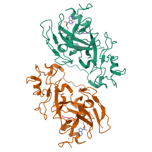
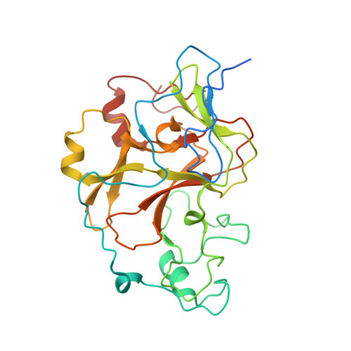
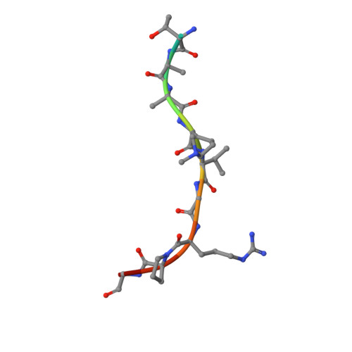
3SWC

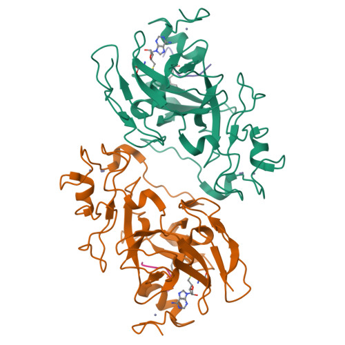
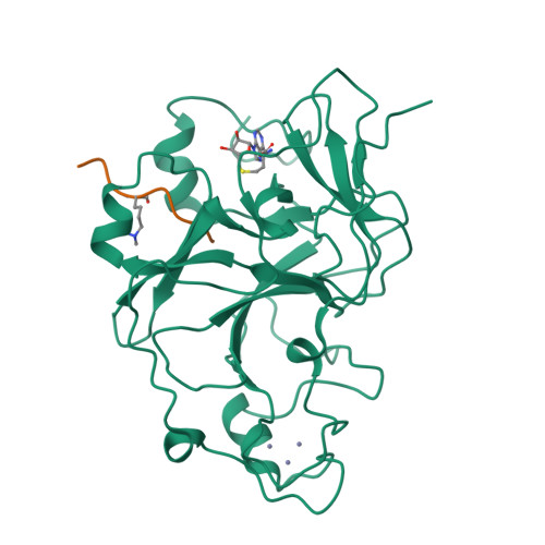
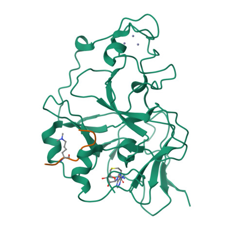
GLP (G9a-like protein) SET domain in complex with Dnmt3aK44me2 peptide


Experimental Data Snapshot

  • Method: X-RAY DIFFRACTION
  • Resolution: 2.33 Å
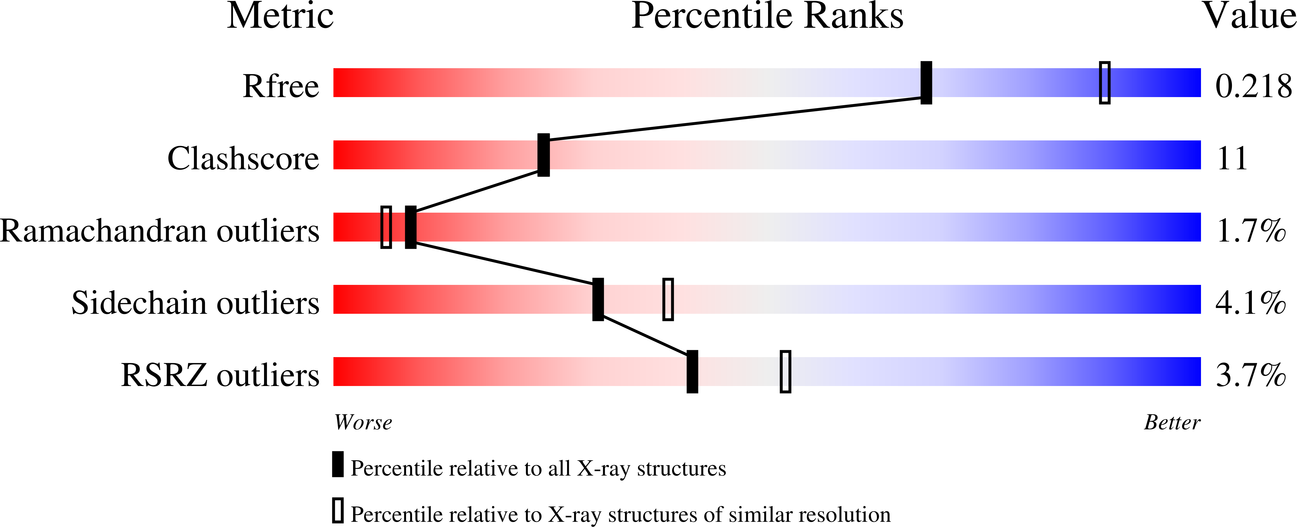
  • R-Value Free: 0.227 
  • R-Value Work: 0.187 
  • R-Value Observed: 0.190 

Starting Model: experimental
View more details

wwPDB Validation   3D Report Full Report


Ligand Structure Quality Assessment 


This is version 1.1 of the entry. See complete history


Literature

MPP8 mediates the interactions between DNA methyltransferase Dnmt3a and H3K9 methyltransferase GLP/G9a.

Chang, Y.Sun, L.Kokura, K.Horton, J.R.Fukuda, M.Espejo, A.Izumi, V.Koomen, J.M.Bedford, M.T.Zhang, X.Shinkai, Y.Fang, J.Cheng, X.

(2011) Nat Commun 2: 533-533

  • DOI: https://doi.org/10.1038/ncomms1549
  • Primary Citation of Related Structures:  
    3SVM, 3SW9, 3SWC

  • PubMed Abstract: 

    DNA CpG methylation and histone H3 lysine 9 (H3K9) methylation are two major repressive epigenetic modifications, and these methylations are positively correlated with one another in chromatin. Here we show that G9a or G9a-like protein (GLP) dimethylate the amino-terminal lysine 44 (K44) of mouse Dnmt3a (equivalent to K47 of human DNMT3A) in vitro and in cells overexpressing G9a or GLP. The chromodomain of MPP8 recognizes the dimethylated Dnmt3aK44me2. MPP8 also interacts with self-methylated GLP in a methylation-dependent manner. The MPP8 chromodomain forms a dimer in solution and in crystals, suggesting that a dimeric MPP8 molecule could bridge the methylated Dnmt3a and GLP, resulting in a silencing complex of Dnmt3a-MPP8-GLP/G9a on chromatin templates. Together, these findings provide a molecular explanation, at least in part, for the co-occurrence of DNA methylation and H3K9 methylation in chromatin.


  • Organizational Affiliation

    Department of Biochemistry, Emory University School of Medicine, Atlanta, Georgia 30322, USA.


Macromolecules
Find similar proteins by:  (by identity cutoff)  |  3D Structure
Entity ID: 1
MoleculeChains Sequence LengthOrganismDetailsImage
Histone-lysine N-methyltransferase EHMT1A,
C [auth B]
285Homo sapiensMutation(s): 0 
Gene Names: EHMT1EUHMTASE1GLPKIAA1876KMT1D
EC: 2.1.1 (PDB Primary Data), 2.1.1.43 (PDB Primary Data), 2.1.1.367 (UniProt)
UniProt & NIH Common Fund Data Resources
Find proteins for Q9H9B1 (Homo sapiens)
Explore Q9H9B1 
Go to UniProtKB:  Q9H9B1
PHAROS:  Q9H9B1
GTEx:  ENSG00000181090 
Entity Groups  
Sequence Clusters30% Identity50% Identity70% Identity90% Identity95% Identity100% Identity
UniProt GroupQ9H9B1
Sequence Annotations
Expand
  • Reference Sequence

Find similar proteins by:  Sequence   |   3D Structure  

Entity ID: 2
MoleculeChains Sequence LengthOrganismDetailsImage
DNA (cytosine-5)-methyltransferase 3AB [auth P],
D [auth Q]
12Mus musculusMutation(s): 0 
EC: 2.1.1.37 (PDB Primary Data), 2.1.1 (UniProt)
UniProt & NIH Common Fund Data Resources
Find proteins for O88508 (Mus musculus)
Explore O88508 
Go to UniProtKB:  O88508
IMPC:  MGI:1261827
Entity Groups  
Sequence Clusters30% Identity50% Identity70% Identity90% Identity95% Identity100% Identity
UniProt GroupO88508
Sequence Annotations
Expand
  • Reference Sequence
Small Molecules
Ligands 2 Unique
IDChains Name / Formula / InChI Key2D Diagram3D Interactions
SAH
Query on SAH

Download Ideal Coordinates CCD File 
I [auth A],
N [auth B]
S-ADENOSYL-L-HOMOCYSTEINE
C14 H20 N6 O5 S
ZJUKTBDSGOFHSH-WFMPWKQPSA-N
ZN
Query on ZN

Download Ideal Coordinates CCD File 
E [auth A]
F [auth A]
G [auth A]
H [auth A]
J [auth B]
E [auth A],
F [auth A],
G [auth A],
H [auth A],
J [auth B],
K [auth B],
L [auth B],
M [auth B]
ZINC ION
Zn
PTFCDOFLOPIGGS-UHFFFAOYSA-N
Modified Residues  1 Unique
IDChains TypeFormula2D DiagramParent
MLY
Query on MLY
B [auth P],
D [auth Q]
L-PEPTIDE LINKINGC8 H18 N2 O2LYS
Binding Affinity Annotations 
IDSourceBinding Affinity
SAH BindingDB:  3SWC IC50: 230 (nM) from 1 assay(s)
Experimental Data & Validation

Experimental Data

  • Method: X-RAY DIFFRACTION
  • Resolution: 2.33 Å
  • R-Value Free: 0.227 
  • R-Value Work: 0.187 
  • R-Value Observed: 0.190 
  • Space Group: P 21 21 21
Unit Cell:
Length ( Å )Angle ( ˚ )
a = 80.09α = 90
b = 90.22β = 90
c = 95.4γ = 90
Software Package:
Software NamePurpose
HKL-2000data collection
PHENIXmodel building
PHENIXrefinement
HKL-2000data reduction
HKL-2000data scaling
PHENIXphasing

Structure Validation

View Full Validation Report


Ligand Structure Quality Assessment 


Entry History 

Deposition Data

Revision History  (Full details and data files)

  • Version 1.0: 2011-11-30
    Type: Initial release
  • Version 1.1: 2023-09-13
    Changes: Data collection, Database references, Derived calculations, Refinement description