3SUB | pdb_00003sub

Crystal structure of the catalytic domain of Plasmodium falciparum ARF GTPase activating protein


Experimental Data Snapshot

  • Method: X-RAY DIFFRACTION
  • Resolution: 2.40 Å
  • R-Value Free: 
    0.227 (Depositor), 0.227 (DCC) 
  • R-Value Work: 
    0.209 (Depositor), 0.206 (DCC) 
  • R-Value Observed: 
    0.209 (Depositor) 

wwPDB Validation 3D Report Full Report

Validation slider image for 3SUB

This is version 1.2 of the entry. See complete history

Literature

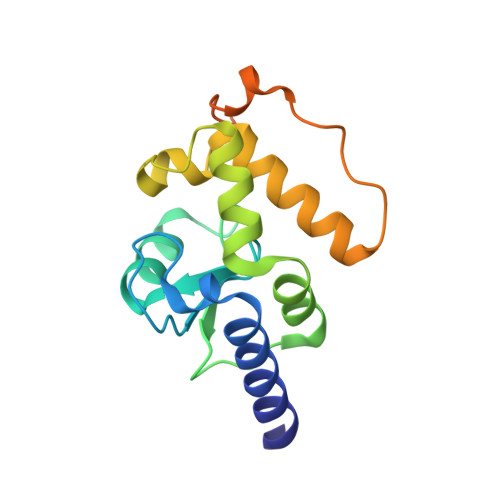
Structure of the catalytic domain of Plasmodium falciparum ARF GTPase-activating protein (ARFGAP).

Cook, W.J.Senkovich, O.Chattopadhyay, D.

(2011) Acta Crystallogr Sect F Struct Biol Cryst Commun 67: 1339-1344

  • DOI: https://doi.org/10.1107/S1744309111032507
  • Primary Citation Related Structures: 
    3SUB

  • PubMed Abstract: 

    The crystal structure of the catalytic domain of the ADP ribosylation factor GTPase-activating protein (ARFGAP) from Plasmodium falciparum has been determined and refined to 2.4 Å resolution. Multiwavength anomalous diffraction (MAD) data were collected utilizing the Zn(2+) ion bound at the zinc-finger domain and were used to solve the structure. The overall structure of the domain is similar to those of mammalian ARFGAPs. However, several amino-acid residues in the area where GAP interacts with ARF1 differ in P. falciparum ARFGAP. Moreover, a number of residues that form the dimer interface in the crystal structure are unique in P. falciparum ARFGAP.


  • Organizational Affiliation
    • Department of Medicine, University of Alabama at Birmingham, CBSE-250, 1015 18th Street South, Birmingham, AL 35294, USA.

Macromolecule Content 

  • Total Structure Weight: 37.56 kDa 
  • Atom Count: 2,299 
  • Modeled Residue Count: 270 
  • Deposited Residue Count: 326 
  • Unique protein chains: 1

Macromolecules

Find similar proteins by:|  3D Structure
Entity ID: 1
MoleculeChains  Sequence LengthOrganismDetailsImage
ADP-ribosylation factor GTPase-activating protein
A, B
163Plasmodium falciparum 3D7Mutation(s): 0 
Gene Names: PFL2140c
UniProt
Find proteins for Q8I4Y5 (Plasmodium falciparum (isolate 3D7))
Explore Q8I4Y5 
Go to UniProtKB:  Q8I4Y5
Entity Groups
Sequence Clusters30% Identity50% Identity70% Identity90% Identity95% Identity100% Identity
UniProt GroupQ8I4Y5
Sequence Annotations
Expand
Reference Sequence

Experimental Data & Validation

Experimental Data

  • Method: X-RAY DIFFRACTION
  • Resolution: 2.40 Å
  • R-Value Free:  0.227 (Depositor), 0.227 (DCC) 
  • R-Value Work:  0.209 (Depositor), 0.206 (DCC) 
  • R-Value Observed: 0.209 (Depositor) 
Space Group: P 32 2 1
Unit Cell:
Length ( Å )Angle ( ˚ )
a = 95.9α = 90
b = 95.9β = 90
c = 92.8γ = 120
Software Package:
Software NamePurpose
HKL-2000data collection
SOLVEphasing
REFMACrefinement
HKL-2000data reduction
HKL-2000data scaling

Structure Validation

View Full Validation Report


Entry History 

Deposition Data

Revision History  (Full details and data files)

  • Version 1.0: 2011-11-09
    Type: Initial release
  • Version 1.1: 2012-03-28
    Changes: Database references
  • Version 1.2: 2024-02-28
    Changes: Data collection, Database references, Derived calculations