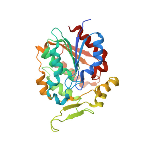
3STV

Crystal Structure of tomato Methylketone Synthase I complexed with 3-hydroxyoctanoate


Experimental Data Snapshot

  • Method: X-RAY DIFFRACTION
  • Resolution: 2.20 Å
  • R-Value Free: 0.236 
  • R-Value Work: 0.202 
  • R-Value Observed: 0.222 

Starting Model: experimental
View more details

wwPDB Validation   3D Report Full Report


Ligand Structure Quality Assessment 


This is version 1.3 of the entry. See complete history


Literature

Emergent Decarboxylase Activity and Attenuation of alpha/beta-Hydrolase Activity during the Evolution of Methylketone Biosynthesis in Tomato.

Auldridge, M.E.Guo, Y.Austin, M.B.Ramsey, J.Fridman, E.Pichersky, E.Noel, J.P.

(2012) Plant Cell 24: 1596-1607

  • DOI: https://doi.org/10.1105/tpc.111.093997
  • Primary Citation of Related Structures:  
    3STT, 3STU, 3STV, 3STW, 3STX, 3STY

  • PubMed Abstract: 

    Specialized methylketone-containing metabolites accumulate in certain plants, in particular wild tomatoes in which they serve as toxic compounds against chewing insects. In Solanum habrochaites f. glabratum, methylketone biosynthesis occurs in the plastids of glandular trichomes and begins with intermediates of de novo fatty acid synthesis. These fatty-acyl intermediates are converted via sequential reactions catalyzed by Methylketone Synthase2 (MKS2) and MKS1 to produce the n-1 methylketone. We report crystal structures of S. habrochaites MKS1, an atypical member of the α/β-hydrolase superfamily. Sequence comparisons revealed the MKS1 catalytic triad, Ala-His-Asn, as divergent to the traditional α/β-hydrolase triad, Ser-His-Asp. Determination of the MKS1 structure points to a novel enzymatic mechanism dependent upon residues Thr-18 and His-243, confirmed by biochemical assays. Structural analysis further reveals a tunnel leading from the active site consisting mostly of hydrophobic residues, an environment well suited for fatty-acyl chain binding. We confirmed the importance of this substrate binding mode by substituting several amino acids leading to an alteration in the acyl-chain length preference of MKS1. Furthermore, we employ structure-guided mutagenesis and functional assays to demonstrate that MKS1, unlike enzymes from this hydrolase superfamily, is not an efficient hydrolase but instead catalyzes the decarboxylation of 3-keto acids.


  • Organizational Affiliation

    Howard Hughes Medical Institute, The Jack H. Skirball Center for Chemical Biology and Proteomics, The Salk Institute for Biological Studies, La Jolla, California 92037, USA. auldridge@wisc.edu


Macromolecules
Find similar proteins by:  (by identity cutoff)  |  3D Structure
Entity ID: 1
MoleculeChains Sequence LengthOrganismDetailsImage
Methylketone synthase 1
A, B
267Solanum habrochaitesMutation(s): 0 
Gene Names: MKS1
UniProt
Find proteins for E0YCS2 (Solanum habrochaites)
Explore E0YCS2 
Go to UniProtKB:  E0YCS2
Entity Groups  
Sequence Clusters30% Identity50% Identity70% Identity90% Identity95% Identity100% Identity
UniProt GroupE0YCS2
Sequence Annotations
Expand
  • Reference Sequence
Small Molecules
Ligands 2 Unique
IDChains Name / Formula / InChI Key2D Diagram3D Interactions
3HO
Query on 3HO

Download Ideal Coordinates CCD File 
C [auth A](3S)-3-hydroxyoctanoic acid
C8 H16 O3
NDPLAKGOSZHTPH-ZETCQYMHSA-N
BR
Query on BR

Download Ideal Coordinates CCD File 
D [auth B]BROMIDE ION
Br
CPELXLSAUQHCOX-UHFFFAOYSA-M
Experimental Data & Validation

Experimental Data

  • Method: X-RAY DIFFRACTION
  • Resolution: 2.20 Å
  • R-Value Free: 0.236 
  • R-Value Work: 0.202 
  • R-Value Observed: 0.222 
  • Space Group: P 1 21 1
Unit Cell:
Length ( Å )Angle ( ˚ )
a = 48.038α = 90
b = 92.38β = 97.56
c = 60.596γ = 90
Software Package:
Software NamePurpose
XSCALEdata scaling
CNSrefinement
PDB_EXTRACTdata extraction
Blu-Icedata collection
XDSdata reduction
CNSphasing

Structure Validation

View Full Validation Report


Ligand Structure Quality Assessment 


Entry History 

Deposition Data

Revision History  (Full details and data files)

  • Version 1.0: 2012-05-02
    Type: Initial release
  • Version 1.1: 2013-06-19
    Changes: Database references
  • Version 1.2: 2017-11-08
    Changes: Refinement description
  • Version 1.3: 2023-09-13
    Changes: Data collection, Database references, Derived calculations, Refinement description