3ST3 | pdb_00003st3

Dreiklang - off state


Experimental Data Snapshot

  • Method: X-RAY DIFFRACTION
  • Resolution: 1.70 Å
  • R-Value Free: 
    0.191 (Depositor), 0.190 (DCC) 
  • R-Value Work: 
    0.159 (Depositor), 0.156 (DCC) 
  • R-Value Observed: 
    0.161 (Depositor) 

wwPDB Validation 3D Report Full Report

Validation slider image for 3ST3

This is version 2.0 of the entry. See complete history

Literature

A reversibly photoswitchable GFP-like protein with fluorescence excitation decoupled from switching.

Brakemann, T.Stiel, A.C.Weber, G.Andresen, M.Testa, I.Grotjohann, T.Leutenegger, M.Plessmann, U.Urlaub, H.Eggeling, C.Wahl, M.C.Hell, S.W.Jakobs, S.

(2011) Nat Biotechnol 29: 942-947

  • DOI: https://doi.org/10.1038/nbt.1952
  • Primary Citation Related Structures: 
    3ST2, 3ST3, 3ST4

  • PubMed Abstract: 

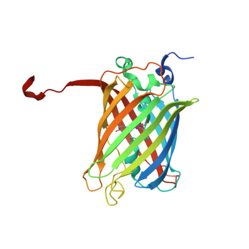
    Photoswitchable fluorescent proteins have enabled new approaches for imaging cells, but their utility has been limited either because they cannot be switched repeatedly or because the wavelengths for switching and fluorescence imaging are strictly coupled. We report a bright, monomeric, reversibly photoswitchable variant of GFP, Dreiklang, whose fluorescence excitation spectrum is decoupled from that for optical switching. Reversible on-and-off switching in living cells is accomplished at illumination wavelengths of ∼365 nm and ∼405 nm, respectively, whereas fluorescence is elicited at ∼515 nm. Mass spectrometry and high-resolution crystallographic analysis of the same protein crystal in the photoswitched on- and off-states demonstrate that switching is based on a reversible hydration/dehydration reaction that modifies the chromophore. The switching properties of Dreiklang enable far-field fluorescence nanoscopy in living mammalian cells using both a coordinate-targeted and a stochastic single molecule switching approach.


  • Organizational Affiliation
    • Max Planck Institute for Biophysical Chemistry, Department of NanoBiophotonics, Göttingen, Germany.

Macromolecule Content 

  • Total Structure Weight: 86.91 kDa 
  • Atom Count: 6,673 
  • Modeled Residue Count: 701 
  • Deposited Residue Count: 750 
  • Unique protein chains: 1

Macromolecules

Find similar proteins by:|  3D Structure
Entity ID: 1
MoleculeChains  Sequence LengthOrganismDetailsImage
Dreiklang
A, B, C
250Aequorea victoriaMutation(s): 0 

Small Molecules

Ligands 1 Unique
IDChains Name / Formula / InChI Key2D Diagram3D Interactions
PO4

Query on PO4


Download:Ideal Coordinates CCD File
D [auth A]
E [auth A]
F [auth A]
G [auth A]
H [auth B]
D [auth A],
E [auth A],
F [auth A],
G [auth A],
H [auth B],
I [auth B],
J [auth B],
K [auth B],
L [auth B],
M [auth C],
N [auth C],
O [auth C],
P [auth C]
PHOSPHATE ION
O4 P
NBIIXXVUZAFLBC-UHFFFAOYSA-K
Modified Residues  1 Unique
IDChains TypeFormula2D DiagramParent
WCR
Query on WCR
A, B, C
L-PEPTIDE LINKINGC13 H15 N3 O5GLY, TYR, GLY

Experimental Data & Validation

Experimental Data

  • Method: X-RAY DIFFRACTION
  • Resolution: 1.70 Å
  • R-Value Free:  0.191 (Depositor), 0.190 (DCC) 
  • R-Value Work:  0.159 (Depositor), 0.156 (DCC) 
  • R-Value Observed: 0.161 (Depositor) 
Space Group: P 21 21 2
Unit Cell:
Length ( Å )Angle ( ˚ )
a = 68.03α = 90
b = 135.34β = 90
c = 81.1γ = 90
Software Package:
Software NamePurpose
MAR345data collection
PHASERphasing
PHENIXrefinement
XDSdata reduction
XDSdata scaling

Structure Validation

View Full Validation Report


Entry History 

Deposition Data

Revision History  (Full details and data files)

  • Version 1.0: 2011-09-14
    Type: Initial release
  • Version 1.1: 2011-09-28
    Changes: Database references
  • Version 1.2: 2011-10-26
    Changes: Database references
  • Version 1.3: 2017-11-08
    Changes: Refinement description
  • Version 1.4: 2024-10-30
    Changes: Data collection, Database references, Derived calculations, Structure summary
  • Version 2.0: 2026-03-18
    Changes: Polymer sequence