


Crystal structure of the Bacillus anthracis acetyl-CoA acetyltransferase
Anderson, S.M., Wawrzak, Z., Onopriyenko, O., Peterson, S.N., Anderson, W.F., Savchenko, A., Center for Structural Genomics of Infectious Diseases (CSGID)To be published.
Experimental Data Snapshot
wwPDB Validation 3D Report Full Report

Entity ID: 1 | |||||
|---|---|---|---|---|---|
| Molecule | Chains | Sequence Length | Organism | Details | Image |
| Acetyl-CoA acetyltransferase | 394 | Bacillus anthracis str. 'Ames Ancestor | Mutation(s): 0 Gene Names: BAS3932, BA_4240, GBAA_4240 EC: 2.3.1.9 | 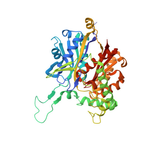 | |
UniProt | |||||
Entity Groups | |||||
| Sequence Clusters | 30% Identity50% Identity70% Identity90% Identity95% Identity100% Identity | ||||
| UniProt Group | A0A6H3AND8 | ||||
Sequence AnnotationsExpand | |||||
Reference Sequence | |||||
| Ligands 2 Unique | |||||
|---|---|---|---|---|---|
| ID | Chains | Name / Formula / InChI Key | 2D Diagram | 3D Interactions | |
| SO4 Download:Ideal Coordinates CCD File | C [auth A] D [auth A] E [auth A] H [auth B] I [auth B] | SULFATE ION O4 S QAOWNCQODCNURD-UHFFFAOYSA-L |  | ||
| K Download:Ideal Coordinates CCD File | F [auth A], G [auth A], L [auth B] | POTASSIUM ION K NPYPAHLBTDXSSS-UHFFFAOYSA-N |  | ||
| Modified Residues 1 Unique | |||||
|---|---|---|---|---|---|
| ID | Chains | Type | Formula | 2D Diagram | Parent |
| CSO Query on CSO | A, B | L-PEPTIDE LINKING | C3 H7 N O3 S |  | CYS |
| Length ( Å ) | Angle ( ˚ ) |
|---|---|
| a = 94.72 | α = 90 |
| b = 94.72 | β = 90 |
| c = 189.26 | γ = 120 |
| Software Name | Purpose |
|---|---|
| PHENIX | refinement |
| PDB_EXTRACT | data extraction |
| BLU-MAX | data collection |
| HKL-2000 | data reduction |
| HKL-2000 | data scaling |
| PHENIX | phasing |


