3SR4

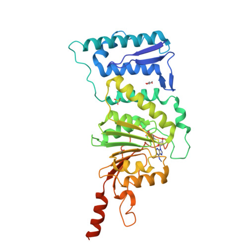
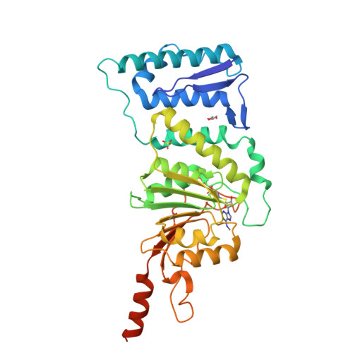
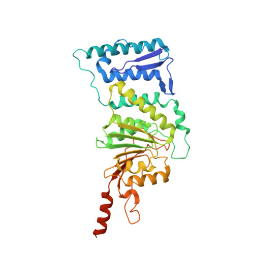
Crystal Structure of Human DOT1L in Complex with a Selective Inhibitor


Experimental Data Snapshot

  • Method: X-RAY DIFFRACTION
  • Resolution: 2.50 Å
  • R-Value Free: 0.274 
  • R-Value Work: 0.234 
  • R-Value Observed: 0.234 

Starting Model: experimental
View more details

wwPDB Validation   3D Report Full Report


Ligand Structure Quality Assessment 


This is version 1.2 of the entry. See complete history


Literature

Selective Inhibitors of Histone Methyltransferase DOT1L: Design, Synthesis, and Crystallographic Studies.

Yao, Y.Chen, P.Diao, J.Cheng, G.Deng, L.Anglin, J.L.Prasad, B.V.Song, Y.

(2011) J Am Chem Soc 133: 16746-16749

  • DOI: https://doi.org/10.1021/ja206312b
  • Primary Citation of Related Structures:  
    3SR4

  • PubMed Abstract: 

    Histone H3-lysine79 (H3K79) methyltransferase DOT1L plays critical roles in normal cell differentiation as well as initiation of acute leukemia. We used structure- and mechanism-based design to discover several potent inhibitors of DOT1L with IC(50) values as low as 38 nM. These inhibitors exhibit only weak or no activities against four other representative histone lysine and arginine methyltransferases, G9a, SUV39H1, PRMT1 and CARM1. The X-ray crystal structure of a DOT1L-inhibitor complex reveals that the N6-methyl group of the inhibitor, located favorably in a predominantly hydrophobic cavity of DOT1L, provides the observed high selectivity. Structural analysis shows that it will disrupt at least one H-bond and/or have steric repulsion for other histone methyltransferases. These compounds represent novel chemical probes for biological function studies of DOT1L in health and disease.


  • Organizational Affiliation

    Department of Pharmacology, Baylor College of Medicine, 1 Baylor Plaza, Houston, Texas 77030, USA.


Macromolecules
Find similar proteins by:  (by identity cutoff)  |  3D Structure
Entity ID: 1
MoleculeChains Sequence LengthOrganismDetailsImage
Histone-lysine N-methyltransferase, H3 lysine-79 specific351Homo sapiensMutation(s): 0 
Gene Names: DOT1LKIAA1814KMT4
EC: 2.1.1.43 (PDB Primary Data), 2.1.1.360 (UniProt)
UniProt & NIH Common Fund Data Resources
Find proteins for Q8TEK3 (Homo sapiens)
Explore Q8TEK3 
Go to UniProtKB:  Q8TEK3
PHAROS:  Q8TEK3
GTEx:  ENSG00000104885 
Entity Groups  
Sequence Clusters30% Identity50% Identity70% Identity90% Identity95% Identity100% Identity
UniProt GroupQ8TEK3
Sequence Annotations
Expand
  • Reference Sequence
Small Molecules
Binding Affinity Annotations 
IDSourceBinding Affinity
TT8 BindingDB:  3SR4 Ki: min: 290, max: 1100 (nM) from 3 assay(s)
PDBBind:  3SR4 Ki: 290 (nM) from 1 assay(s)
Experimental Data & Validation

Experimental Data

  • Method: X-RAY DIFFRACTION
  • Resolution: 2.50 Å
  • R-Value Free: 0.274 
  • R-Value Work: 0.234 
  • R-Value Observed: 0.234 
  • Space Group: P 65
Unit Cell:
Length ( Å )Angle ( ˚ )
a = 152.753α = 90
b = 152.753β = 90
c = 50.889γ = 120
Software Package:
Software NamePurpose
StructureStudiodata collection
PHASERphasing
CNSrefinement
HKL-2000data reduction
HKL-2000data scaling

Structure Validation

View Full Validation Report


Ligand Structure Quality Assessment 


Entry History 

Deposition Data

Revision History  (Full details and data files)

  • Version 1.0: 2011-10-05
    Type: Initial release
  • Version 1.1: 2011-11-16
    Changes: Database references
  • Version 1.2: 2023-09-13
    Changes: Data collection, Database references, Derived calculations, Refinement description