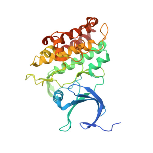
Crystal structure of Activin receptor type-IIA (ACVR2A) kinase domain in complex with a quinazolin
Chaikuad, A., Williams, E., Mahajan, P., Cooper, C.D.O., Sanvitale, C., Vollmar, M., Muniz, J.R.C., Yue, W.W., von Delft, F., Weigelt, J., Arrowsmith, C.H., Edwards, A.M., Bountra, C., Bullock, A., Structural Genomics Consortium (SGC)To be published.