


Crystal structure of human CYP7A1 in complex with cholest-4-en-3-one
Strushkevich, N., Tempel, W., MacKenzie, F., Wernimont, A.K., Arrowsmith, C.H., Edwards, A.M., Bountra, C., Weigelt, J., Park, H.To be published.
Experimental Data Snapshot
Starting Model: experimental
View more details
Entity ID: 1 | |||||
|---|---|---|---|---|---|
| Molecule | Chains | Sequence Length | Organism | Details | Image |
| Cholesterol 7-alpha-monooxygenase | 491 | Homo sapiens | Mutation(s): 1 Gene Names: CYP7A1, CYP7 EC: 1.14.13.17 (PDB Primary Data), 1.14.14.26 (UniProt), 1.14.14.23 (UniProt) Membrane Entity: Yes | 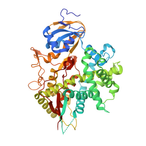 | |
UniProt & NIH Common Fund Data Resources | |||||
PHAROS: P22680 GTEx: ENSG00000167910 | |||||
Entity Groups | |||||
| Sequence Clusters | 30% Identity50% Identity70% Identity90% Identity95% Identity100% Identity | ||||
| UniProt Group | P22680 | ||||
Sequence AnnotationsExpand | |||||
Reference Sequence | |||||
| Ligands 3 Unique | |||||
|---|---|---|---|---|---|
| ID | Chains | Name / Formula / InChI Key | 2D Diagram | 3D Interactions | |
| HEM Download:Ideal Coordinates CCD File | C [auth A], I [auth B] | PROTOPORPHYRIN IX CONTAINING FE C34 H32 Fe N4 O4 KABFMIBPWCXCRK-RGGAHWMASA-L |  | ||
| K2B Download:Ideal Coordinates CCD File | D [auth A], J [auth B] | (8ALPHA,9BETA)-CHOLEST-4-EN-3-ONE C27 H44 O NYOXRYYXRWJDKP-GYKMGIIDSA-N |  | ||
| UNX Download:Ideal Coordinates CCD File | E [auth A] F [auth A] G [auth A] H [auth A] K [auth B] | UNKNOWN ATOM OR ION X |  | ||
| Length ( Å ) | Angle ( ˚ ) |
|---|---|
| a = 56.16 | α = 90 |
| b = 137.63 | β = 90 |
| c = 160.15 | γ = 90 |
| Software Name | Purpose |
|---|---|
| XSCALE | data scaling |
| PHASER | phasing |
| BUSTER-TNT | refinement |
| PDB_EXTRACT | data extraction |
| Blu-Ice | data collection |
| XDS | data reduction |
| BUSTER | refinement |


