3SMQ | pdb_00003smq

Crystal structure of protein arginine methyltransferase 3


Experimental Data Snapshot

  • Method: X-RAY DIFFRACTION
  • Resolution: 2.00 Å
  • R-Value Free: 
    0.215 (Depositor), 0.220 (DCC) 
  • R-Value Work: 
    0.190 (Depositor), 0.191 (DCC) 
  • R-Value Observed: 
    0.191 (Depositor) 

Starting Model: experimental
View more details

wwPDB Validation 3D Report Full Report

Validation slider image for 3SMQ

Ligand Structure Quality Assessment 


This is version 1.2 of the entry. See complete history

Literature

An allosteric inhibitor of protein arginine methyltransferase 3.

Siarheyeva, A.Senisterra, G.Allali-Hassani, A.Dong, A.Dobrovetsky, E.Wasney, G.A.Chau, I.Marcellus, R.Hajian, T.Liu, F.Korboukh, I.Smil, D.Bolshan, Y.Min, J.Wu, H.Zeng, H.Loppnau, P.Poda, G.Griffin, C.Aman, A.Brown, P.J.Jin, J.Al-Awar, R.Arrowsmith, C.H.Schapira, M.Vedadi, M.

(2012) Structure 20: 1425-1435

  • DOI: https://doi.org/10.1016/j.str.2012.06.001
  • Primary Citation Related Structures: 
    3SMQ

  • PubMed Abstract: 

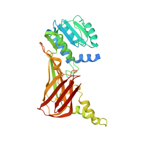
    PRMT3, a protein arginine methyltransferase, has been shown to influence ribosomal biosynthesis by catalyzing the dimethylation of the 40S ribosomal protein S2. Although PRMT3 has been reported to be a cytosolic protein, it has been shown to methylate histone H4 peptide (H4 1-24) in vitro. Here, we report the identification of a PRMT3 inhibitor (1-(benzo[d][1,2,3]thiadiazol-6-yl)-3-(2-cyclohexenylethyl)urea; compound 1) with IC50 value of 2.5 μM by screening a library of 16,000 compounds using H4 (1-24) peptide as a substrate. The crystal structure of PRMT3 in complex with compound 1 as well as kinetic analysis reveals an allosteric mechanism of inhibition. Mutating PRMT3 residues within the allosteric site or using compound 1 analogs that disrupt interactions with allosteric site residues both abrogated binding and inhibitory activity. These data demonstrate an allosteric mechanism for inhibition of protein arginine methyltransferases, an emerging class of therapeutic targets.


  • Organizational Affiliation
    • Structural Genomics Consortium, University of Toronto, 101 College Street, MaRS Centre, South Tower, Toronto, ON M5G 1L7, Canada.

Macromolecule Content 

  • Total Structure Weight: 38.62 kDa 
  • Atom Count: 2,632 
  • Modeled Residue Count: 299 
  • Deposited Residue Count: 340 
  • Unique protein chains: 1

Macromolecules

Find similar proteins by:|  3D Structure
Entity ID: 1
MoleculeChains  Sequence LengthOrganismDetailsImage
Protein arginine N-methyltransferase 3340Homo sapiensMutation(s): 1 
Gene Names: PRMT3HRMT1L3
EC: 2.1.1 (PDB Primary Data), 2.1.1.319 (UniProt)
UniProt & NIH Common Fund Data Resources
Find proteins for O60678 (Homo sapiens)
Explore O60678 
Go to UniProtKB:  O60678
PHAROS:  O60678
GTEx:  ENSG00000185238 
Entity Groups
Sequence Clusters30% Identity50% Identity70% Identity90% Identity95% Identity100% Identity
UniProt GroupO60678
Sequence Annotations
Expand
Reference Sequence

Small Molecules

Ligands 3 Unique
IDChains Name / Formula / InChI Key2D Diagram3D Interactions
TDU

Query on TDU


Download:Ideal Coordinates CCD File
B [auth A]1-(1,2,3-benzothiadiazol-6-yl)-3-[2-(cyclohex-1-en-1-yl)ethyl]urea
C15 H18 N4 O S
GGXCUZHEJUJACD-UHFFFAOYSA-N
CL

Query on CL


Download:Ideal Coordinates CCD File
C [auth A]CHLORIDE ION
Cl
VEXZGXHMUGYJMC-UHFFFAOYSA-M
UNX

Query on UNX


Download:Ideal Coordinates CCD File
D [auth A]
E [auth A]
F [auth A]
G [auth A]
H [auth A]
D [auth A],
E [auth A],
F [auth A],
G [auth A],
H [auth A],
I [auth A],
J [auth A],
K [auth A],
L [auth A],
M [auth A]
UNKNOWN ATOM OR ION
X
Binding Affinity Annotations 
IDSourceBinding Affinity
TDU BindingDB:  3SMQ Kd: 9500 (nM) from 1 assay(s)
IC50: min: 1600, max: 2500 (nM) from 3 assay(s)

Experimental Data & Validation

Experimental Data

  • Method: X-RAY DIFFRACTION
  • Resolution: 2.00 Å
  • R-Value Free:  0.215 (Depositor), 0.220 (DCC) 
  • R-Value Work:  0.190 (Depositor), 0.191 (DCC) 
  • R-Value Observed: 0.191 (Depositor) 
Space Group: P 43 21 2
Unit Cell:
Length ( Å )Angle ( ˚ )
a = 70.649α = 90
b = 70.649β = 90
c = 171.977γ = 90
Software Package:
Software NamePurpose
MOLREPphasing
REFMACrefinement
Cootmodel building
HKL-2000data reduction
HKL-2000data scaling

Structure Validation

View Full Validation Report


Ligand Structure Quality Assessment 


Entry History 

Revision History  (Full details and data files)

  • Version 1.0: 2011-08-31
    Type: Initial release
  • Version 1.1: 2013-07-24
    Changes: Database references
  • Version 1.2: 2023-09-13
    Changes: Data collection, Database references, Derived calculations, Refinement description