3SKN | pdb_00003skn

Crystal structure of the RL42 TCR unliganded


Experimental Data Snapshot

  • Method: X-RAY DIFFRACTION
  • Resolution: 2.90 Å
  • R-Value Free: 
    0.292 (Depositor), 0.278 (DCC) 
  • R-Value Work: 
    0.194 (Depositor) 
  • R-Value Observed: 
    0.199 (Depositor) 

Starting Model: experimental
View more details

wwPDB Validation 3D Report Full Report

Validation slider image for 3SKN

This is version 1.3 of the entry. See complete history

Literature

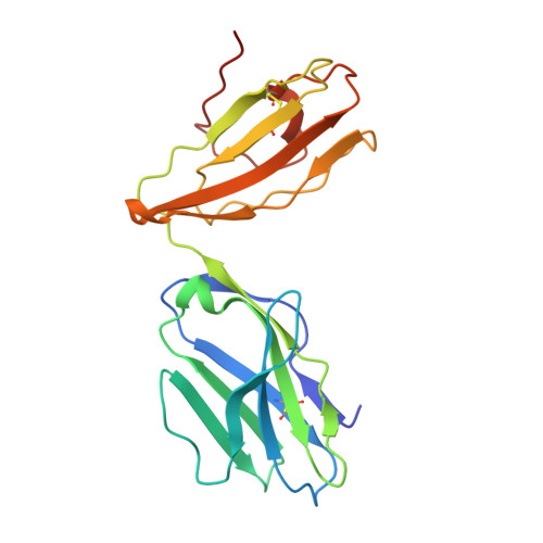
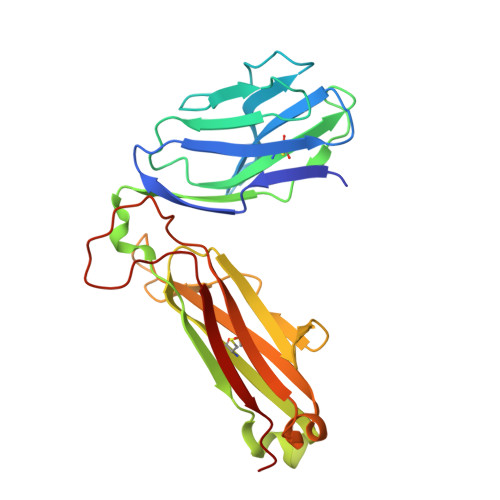
A structural basis for varied alpha-beta TCR usage against an immunodominant EBV antigen restricted to a HLA-B8 molecule.

Gras, S.Wilmann, P.G.Chen, Z.Halim, H.Liu, Y.C.Kjer-Nielsen, L.Purcell, A.W.Burrows, S.R.McCluskey, J.Rossjohn, J.

(2012) J Immunol 188: 311-321

  • DOI: https://doi.org/10.4049/jimmunol.1102686
  • Primary Citation Related Structures: 
    3SJV, 3SKM, 3SKN, 3SKO

  • PubMed Abstract: 

    EBV is a ubiquitous and persistent human pathogen, kept in check by the cytotoxic T cell response. In this study, we investigated how three TCRs, which differ in their T cell immunodominance hierarchies and gene usage, interact with the same EBV determinant (FLRGRAYGL), bound to the same Ag-presenting molecule, HLA-B8. We found that the three TCRs exhibit differing fine specificities for the viral Ag. Further, via structural and biophysical approaches, we demonstrated that the viral Ag provides the greatest energetic contribution to the TCR-peptide-HLA interaction, while focusing on a few adjacent HLA-based interactions to further tune fine-specificity requirements. Thus, the TCR engages the peptide-HLA with the viral Ag as the main glue, such that neighboring TCR-MHC interactions are recruited as a supportive adhesive. Collectively, we provide a portrait of how the host's adaptive immune response differentially engages a common viral Ag.


  • Organizational Affiliation
    • Department of Biochemistry and Molecular Biology, Monash University, Clayton, Victoria, Australia.

Macromolecule Content 

  • Total Structure Weight: 200.6 kDa 
  • Atom Count: 13,825 
  • Modeled Residue Count: 1,752 
  • Deposited Residue Count: 1,788 
  • Unique protein chains: 2

Macromolecules

Find similar proteins by:|  3D Structure
Entity ID: 1
MoleculeChains  Sequence LengthOrganismDetailsImage
RL42 T cell receptor, alpha chain
A, C, E, G
203Homo sapiensMutation(s): 0 
UniProt & NIH Common Fund Data Resources
Find proteins for P01848 (Homo sapiens)
Explore P01848 
Go to UniProtKB:  P01848
PHAROS:  P01848
Entity Groups
Sequence Clusters30% Identity50% Identity70% Identity90% Identity95% Identity100% Identity
UniProt GroupP01848
Sequence Annotations
Expand
Reference Sequence
Find similar proteins by:|  3D Structure
Entity ID: 2
MoleculeChains  Sequence LengthOrganismDetailsImage
RL42 T cell receptor, beta chain
B, D, F, H
244Homo sapiensMutation(s): 0 

Experimental Data & Validation

Experimental Data

  • Method: X-RAY DIFFRACTION
  • Resolution: 2.90 Å
  • R-Value Free:  0.292 (Depositor), 0.278 (DCC) 
  • R-Value Work:  0.194 (Depositor) 
  • R-Value Observed: 0.199 (Depositor) 
Space Group: P 1
Unit Cell:
Length ( Å )Angle ( ˚ )
a = 74.97α = 69.35
b = 77.81β = 72.2
c = 99.64γ = 83.33
Software Package:
Software NamePurpose
XSCALEdata scaling
PHASERphasing
PHENIXrefinement
PDB_EXTRACTdata extraction
Blu-Icedata collection

Structure Validation

View Full Validation Report


Entry History 

Deposition Data

Revision History  (Full details and data files)

  • Version 1.0: 2012-02-29
    Type: Initial release
  • Version 1.1: 2012-08-01
    Changes: Structure summary
  • Version 1.2: 2023-09-13
    Changes: Data collection, Database references, Refinement description
  • Version 1.3: 2024-11-06
    Changes: Structure summary