3SIP | pdb_00003sip

Crystal structure of drICE and dIAP1-BIR1 complex


Experimental Data Snapshot

  • Method: X-RAY DIFFRACTION
  • Resolution: 3.50 Å
  • R-Value Free: 
    0.256 (Depositor), 0.257 (DCC) 
  • R-Value Work: 
    0.210 (Depositor), 0.207 (DCC) 
  • R-Value Observed: 
    0.212 (Depositor) 

Starting Model: experimental
View more details

wwPDB Validation 3D Report Full Report

Validation slider image for 3SIP

This is version 1.4 of the entry. See complete history

Literature

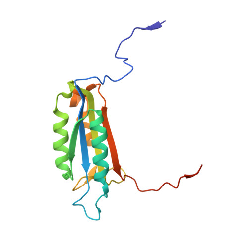
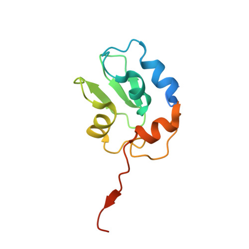
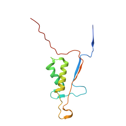
Structural mechanisms of DIAP1 auto-inhibition and DIAP1-mediated inhibition of drICE.

Li, X.Wang, J.Shi, Y.

(2011) Nat Commun 2: 408-408

  • DOI: https://doi.org/10.1038/ncomms1418
  • Primary Citation Related Structures: 
    3SIP, 3SIQ, 3SIR

  • PubMed Abstract: 

    The Drosophila inhibitor of apoptosis protein DIAP1 exists in an auto-inhibited conformation, unable to suppress the effector caspase drICE. Auto-inhibition is disabled by caspase-mediated cleavage of DIAP1 after Asp20. The cleaved DIAP1 binds to mature drICE, inhibits its protease activity, and, presumably, also targets drICE for ubiquitylation. DIAP1-mediated suppression of drICE is effectively antagonized by the pro-apoptotic proteins Reaper, Hid, and Grim (RHG). Despite rigorous effort, the molecular mechanisms behind these observations are enigmatic. Here we report a 2.4 Å crystal structure of uncleaved DIAP1-BIR1, which reveals how the amino-terminal sequences recognize a conserved surface groove in BIR1 to achieve auto-inhibition, and a 3.5 Å crystal structure of active drICE bound to cleaved DIAP1-BIR1, which provides a structural explanation to DIAP1-mediated inhibition of drICE. These structures and associated biochemical analyses, together with published reports, define the molecular determinants that govern the interplay among DIAP1, drICE and the RHG proteins.


  • Organizational Affiliation
    • Ministry of Education Protein Science Laboratory, Center for Structural Biology, School of Life Sciences and School of Medicine, Tsinghua University, Beijing, China.

Macromolecule Content 

  • Total Structure Weight: 87.4 kDa 
  • Atom Count: 5,714 
  • Modeled Residue Count: 712 
  • Deposited Residue Count: 762 
  • Unique protein chains: 3

Macromolecules

Find similar proteins by:|  3D Structure
Entity ID: 1
MoleculeChains  Sequence LengthOrganismDetailsImage
CaspaseA,
B [auth C]
157Drosophila melanogasterMutation(s): 0 
EC: 3.4.22
UniProt
Find proteins for O01382 (Drosophila melanogaster)
Explore O01382 
Go to UniProtKB:  O01382
Entity Groups
Sequence Clusters30% Identity50% Identity70% Identity90% Identity95% Identity100% Identity
UniProt GroupO01382
Sequence Annotations
Expand
Reference Sequence
Find similar proteins by:|  3D Structure
Entity ID: 2
MoleculeChains  Sequence LengthOrganismDetailsImage
Apoptosis 1 inhibitorC [auth E],
D [auth F]
115Drosophila melanogasterMutation(s): 1 
EC: 6.3.2 (PDB Primary Data), 2.3.2.27 (UniProt)
UniProt
Find proteins for Q24306 (Drosophila melanogaster)
Explore Q24306 
Go to UniProtKB:  Q24306
Entity Groups
Sequence Clusters30% Identity50% Identity70% Identity90% Identity95% Identity100% Identity
UniProt GroupQ24306
Sequence Annotations
Expand
Reference Sequence
Find similar proteins by:|  3D Structure
Entity ID: 3
MoleculeChains  Sequence LengthOrganismDetailsImage
CaspaseE [auth B],
F [auth D]
109Drosophila melanogasterMutation(s): 0 
EC: 3.4.22
UniProt
Find proteins for O01382 (Drosophila melanogaster)
Explore O01382 
Go to UniProtKB:  O01382
Entity Groups
Sequence Clusters30% Identity50% Identity70% Identity90% Identity95% Identity100% Identity
UniProt GroupO01382
Sequence Annotations
Expand
Reference Sequence

Experimental Data & Validation

Experimental Data

  • Method: X-RAY DIFFRACTION
  • Resolution: 3.50 Å
  • R-Value Free:  0.256 (Depositor), 0.257 (DCC) 
  • R-Value Work:  0.210 (Depositor), 0.207 (DCC) 
  • R-Value Observed: 0.212 (Depositor) 
Space Group: I 21 21 21
Unit Cell:
Length ( Å )Angle ( ˚ )
a = 91.35α = 90
b = 148.115β = 90
c = 156.738γ = 90
Software Package:
Software NamePurpose
PHENIXrefinement

Structure Validation

View Full Validation Report


Entry History 

Deposition Data

Revision History  (Full details and data files)

  • Version 1.0: 2011-08-10
    Type: Initial release
  • Version 1.1: 2013-07-10
    Changes: Database references
  • Version 1.2: 2014-05-21
    Changes: Other
  • Version 1.3: 2018-07-25
    Changes: Data collection, Source and taxonomy, Structure summary
  • Version 1.4: 2023-11-01
    Changes: Data collection, Database references, Derived calculations, Refinement description