3SFM | pdb_00003sfm

Novel crystallization conditions for tandem variant R67 DHFR yields wild-type crystal structure


Experimental Data Snapshot

  • Method: X-RAY DIFFRACTION
  • Resolution: 1.40 Å
  • R-Value Free: 
    0.164 (Depositor), 0.156 (DCC) 
  • R-Value Work: 
    0.144 (Depositor), 0.136 (DCC) 
  • R-Value Observed: 
    0.145 (Depositor) 

Starting Model: experimental
View more details

wwPDB Validation 3D Report Full Report

Validation slider image for 3SFM

This is version 1.2 of the entry. See complete history

Literature

Novel crystallization conditions for tandem variant R67 DHFR yield a wild-type crystal structure.

Yachnin, B.J.Colin, D.Y.Volpato, J.P.Ebert, M.Pelletier, J.N.Berghuis, A.M.

(2011) Acta Crystallogr Sect F Struct Biol Cryst Commun 67: 1316-1322

  • DOI: https://doi.org/10.1107/S1744309111030417
  • Primary Citation Related Structures: 
    3SFM

  • PubMed Abstract: 

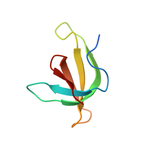
    Trimethoprim is an antibiotic that targets bacterial dihydrofolate reductase (DHFR). A plasmid-encoded DHFR known as R67 DHFR provides resistance to trimethoprim in bacteria. To better understand the mechanism of this homotetrameric enzyme, a tandem dimer construct was created that linked two monomeric R67 DHFR subunits together and mutated the sequence of residues 66-69 of the first subunit from VQIY to INSF. Using a modified crystallization protocol for this enzyme that included in situ proteolysis using chymotrypsin, the tandem dimer was crystallized and the structure was solved at 1.4 Å resolution. Surprisingly, only wild-type protomers were incorporated into the crystal. Further experiments demonstrated that the variant protomer was selectively degraded by chymotrypsin, although no canonical chymotrypsin cleavage site had been introduced by these mutations.


  • Organizational Affiliation
    • Department of Biochemistry, McGill University, 3649 Promenade Sir William Osler, Bellini Pavilion, Room 470, Montreal, Quebec H3G 0B1, Canada.

Macromolecule Content 

  • Total Structure Weight: 7.09 kDa 
  • Atom Count: 567 
  • Modeled Residue Count: 58 
  • Deposited Residue Count: 62 
  • Unique protein chains: 1

Macromolecules

Find similar proteins by:|  3D Structure
Entity ID: 1
MoleculeChains  Sequence LengthOrganismDetailsImage
Dihydrofolate reductase type 262Escherichia coliMutation(s): 1 
Gene Names: dfrB
EC: 1.5.1.3
UniProt
Find proteins for P00383 (Escherichia coli)
Explore P00383 
Go to UniProtKB:  P00383
Entity Groups
Sequence Clusters30% Identity50% Identity70% Identity90% Identity95% Identity100% Identity
UniProt GroupP00383
Sequence Annotations
Expand
Reference Sequence

Experimental Data & Validation

Experimental Data

  • Method: X-RAY DIFFRACTION
  • Resolution: 1.40 Å
  • R-Value Free:  0.164 (Depositor), 0.156 (DCC) 
  • R-Value Work:  0.144 (Depositor), 0.136 (DCC) 
  • R-Value Observed: 0.145 (Depositor) 
Space Group: I 41 2 2
Unit Cell:
Length ( Å )Angle ( ˚ )
a = 67.691α = 90
b = 67.691β = 90
c = 51.76γ = 90
Software Package:
Software NamePurpose
StructureStudiodata collection
REFMACrefinement
HKL-2000data reduction
HKL-2000data scaling
REFMACphasing

Structure Validation

View Full Validation Report


Entry History 

Deposition Data

Revision History  (Full details and data files)

  • Version 1.0: 2011-11-02
    Type: Initial release
  • Version 1.1: 2011-12-07
    Changes: Database references
  • Version 1.2: 2023-09-13
    Changes: Data collection, Database references, Derived calculations, Refinement description