3SFG | pdb_00003sfg

crystal structure of murine norovirus RNA dependent RNA polymerase in complex with 2thiouridine(2TU)


Experimental Data Snapshot

  • Method: X-RAY DIFFRACTION
  • Resolution: 2.21 Å
  • R-Value Free: 
    0.253 (Depositor), 0.224 (DCC) 
  • R-Value Work: 
    0.203 (Depositor) 
  • R-Value Observed: 
    0.203 (Depositor) 

wwPDB Validation 3D Report Full Report

Validation slider image for 3SFG

Ligand Structure Quality Assessment 


This is version 1.1 of the entry. See complete history

Literature

Crystal structures of murine norovirus-1 RNA-dependent RNA polymerase in complex with 2-thiouridine or ribavirin.

Alam, I.Lee, J.H.Cho, K.J.Han, K.R.Yang, J.M.Chung, M.S.Kim, K.H.

(2012) Virology 426: 143-151

  • DOI: https://doi.org/10.1016/j.virol.2012.01.016
  • Primary Citation Related Structures: 
    3SFG, 3SFU

  • PubMed Abstract: 

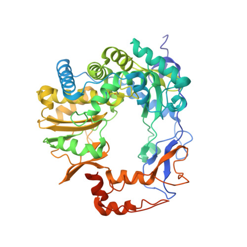
    Murine norovirus-1 (MNV-1) shares many features with human norovirus (HuNoV) and both are classified within the norovirus genus of Caliciviridae family. MNV-1 is used as the surrogate for HuNoV research since it is the only form that can be grown in cell culture. HuNoV and MNV-1 RNA dependent RNA polymerase (RdRp) proteins with the sequence identity of 59% show essentially identical conformations. Here we report the first structural evidence of 2-thiouridine (2TU) or ribavirin binding to MNV-1 RdRp, based on the crystal structures determined at 2.2Å and 2.5Å resolutions, respectively. Cellular and biochemical studies revealed stronger inhibitory effect of 2TU on the replication of MNV-1 in RAW 264.7 cells, compared to that of ribavirin. Our complex structures highlight the key interactions involved in recognition of the nucleoside analogs which block the active site of the viral RNA polymerase.


  • Organizational Affiliation
    • Department of Biotechnology & Bioinformatics, Korea University, Chungnam 339-700, Republic of Korea.

Macromolecule Content 

  • Total Structure Weight: 181.4 kDa 
  • Atom Count: 12,359 
  • Modeled Residue Count: 1,442 
  • Deposited Residue Count: 1,551 
  • Unique protein chains: 1

Macromolecules

Find similar proteins by:|  3D Structure
Entity ID: 1
MoleculeChains  Sequence LengthOrganismDetailsImage
RNA polymerase
A, B, C
517Murine norovirus 1Mutation(s): 0 
Gene Names: RdRp
UniProt
Find proteins for Q80J95 (Norovirus (isolate Mouse/NoV/United States/MNV1/2002/GV))
Explore Q80J95 
Go to UniProtKB:  Q80J95
Entity Groups
Sequence Clusters30% Identity50% Identity70% Identity90% Identity95% Identity100% Identity
UniProt GroupQ80J95
Sequence Annotations
Expand
Reference Sequence

Small Molecules

Ligands 4 Unique
IDChains Name / Formula / InChI Key2D Diagram3D Interactions
2TU

Query on 2TU


Download:Ideal Coordinates CCD File
AA [auth B],
D [auth A],
RA [auth C]
1-(beta-D-ribofuranosyl)-2-thioxo-2,3-dihydropyrimidin-4(1H)-one
C9 H12 N2 O5 S
GJTBSTBJLVYKAU-XVFCMESISA-N
SO4

Query on SO4


Download:Ideal Coordinates CCD File
AB [auth C]
BA [auth B]
BB [auth C]
CA [auth B]
CB [auth C]
AB [auth C],
BA [auth B],
BB [auth C],
CA [auth B],
CB [auth C],
DA [auth B],
E [auth A],
EA [auth B],
F [auth A],
FA [auth B],
G [auth A],
GA [auth B],
H [auth A],
HA [auth B],
I [auth A],
IA [auth B],
J [auth A],
K [auth A],
L [auth A],
M [auth A],
SA [auth C],
TA [auth C],
UA [auth C],
VA [auth C],
WA [auth C],
XA [auth C],
YA [auth C],
ZA [auth C]
SULFATE ION
O4 S
QAOWNCQODCNURD-UHFFFAOYSA-L
GOL

Query on GOL


Download:Ideal Coordinates CCD File
DB [auth C]
EB [auth C]
FB [auth C]
GB [auth C]
HB [auth C]
DB [auth C],
EB [auth C],
FB [auth C],
GB [auth C],
HB [auth C],
IB [auth C],
JA [auth B],
KA [auth B],
LA [auth B],
MA [auth B],
N [auth A],
NA [auth B],
O [auth A],
OA [auth B],
P [auth A],
PA [auth B],
Q [auth A],
R [auth A],
S [auth A],
T [auth A],
U [auth A],
V [auth A],
W [auth A],
X [auth A],
Y [auth A]
GLYCEROL
C3 H8 O3
PEDCQBHIVMGVHV-UHFFFAOYSA-N
MG

Query on MG


Download:Ideal Coordinates CCD File
JB [auth C],
QA [auth B],
Z [auth A]
MAGNESIUM ION
Mg
JLVVSXFLKOJNIY-UHFFFAOYSA-N

Experimental Data & Validation

Experimental Data

  • Method: X-RAY DIFFRACTION
  • Resolution: 2.21 Å
  • R-Value Free:  0.253 (Depositor), 0.224 (DCC) 
  • R-Value Work:  0.203 (Depositor) 
  • R-Value Observed: 0.203 (Depositor) 
Space Group: C 1 2 1
Unit Cell:
Length ( Å )Angle ( ˚ )
a = 120.794α = 90
b = 195.972β = 114.86
c = 109.335γ = 90
Software Package:
Software NamePurpose
PHENIXrefinement
PDB_EXTRACTdata extraction

Structure Validation

View Full Validation Report


Ligand Structure Quality Assessment 


Entry History 

Deposition Data

Revision History  (Full details and data files)

  • Version 1.0: 2012-05-09
    Type: Initial release
  • Version 1.1: 2024-10-30
    Changes: Data collection, Database references, Derived calculations, Structure summary