3S7X

Unassembled Washington University Polyomavirus VP1 Pentamer R198K Mutant


Experimental Data Snapshot

  • Method: X-RAY DIFFRACTION
  • Resolution: 2.90 Å
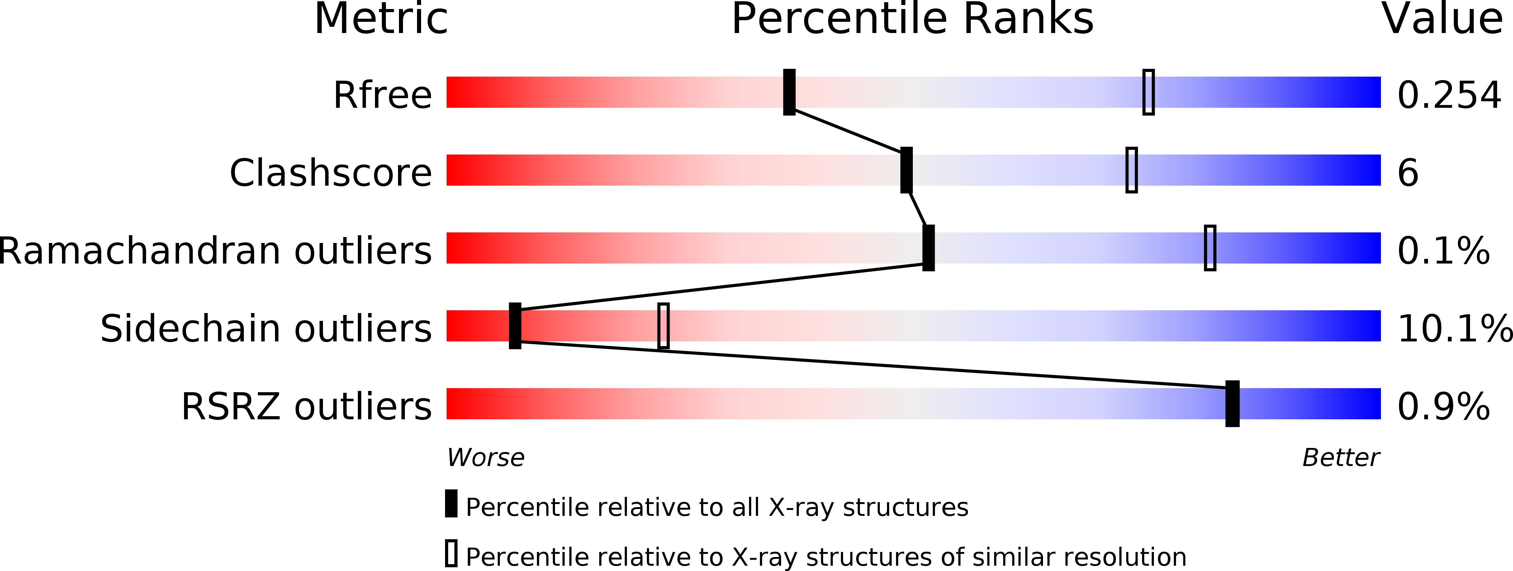
  • R-Value Free: 0.253 
  • R-Value Work: 0.234 
  • R-Value Observed: 0.235 

wwPDB Validation   3D Report Full Report


This is version 1.1 of the entry. See complete history


Literature

Structures of the major capsid proteins of the human KI and WU polyomaviruses.

Neu, U.Wang, J.Macejak, D.Garcea, R.L.Stehle, T.

(2011) J Virol 85: 7384-7392

  • DOI: https://doi.org/10.1128/JVI.00382-11
  • Primary Citation of Related Structures:  
    3S7V, 3S7X

  • PubMed Abstract: 

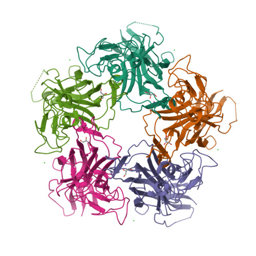
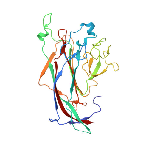
    The Karolinska Institutet and Washington University polyomaviruses (KIPyV and WUPyV, respectively) are recently discovered human viruses that infect the respiratory tract. Although they have not yet been linked to disease, they are prevalent in populations worldwide, with initial infection occurring in early childhood. Polyomavirus capsids consist of 72 pentamers of the major capsid protein viral protein 1 (VP1), which determines antigenicity and receptor specificity. The WUPyV and KIPyV VP1 proteins are distant in evolution from VP1 proteins of known structure such as simian virus 40 or murine polyomavirus. We present here the crystal structures of unassembled recombinant WUPyV and KIPyV VP1 pentamers at resolutions of 2.9 and 2.55 Å, respectively. The WUPyV and KIPyV VP1 core structures fold into the same β-sandwich that is a hallmark of all polyomavirus VP1 proteins crystallized to date. However, differences in sequence translate into profoundly different surface loop structures in KIPyV and WUPyV VP1 proteins. Such loop structures have not been observed for other polyomaviruses, and they provide initial clues about the possible interactions of these viruses with cell surface receptors.


  • Organizational Affiliation

    Interfaculty Institute of Biochemistry, University of Tuebingen, Hoppe-Seyler-Str. 4, D-72076 Tuebingen, Germany.


Macromolecules
Find similar proteins by:  (by identity cutoff)  |  3D Structure
Entity ID: 1
MoleculeChains Sequence LengthOrganismDetailsImage
Major capsid protein VP1
A, B, C, D, E
267WU PolyomavirusMutation(s): 1 
Gene Names: VP1
UniProt
Find proteins for A5HBD5 (WU polyomavirus)
Explore A5HBD5 
Go to UniProtKB:  A5HBD5
Entity Groups  
Sequence Clusters30% Identity50% Identity70% Identity90% Identity95% Identity100% Identity
UniProt GroupA5HBD5
Sequence Annotations
Expand
  • Reference Sequence
Small Molecules
Ligands 3 Unique
IDChains Name / Formula / InChI Key2D Diagram3D Interactions
GOL
Query on GOL

Download Ideal Coordinates CCD File 
F [auth A],
K [auth B],
M [auth C],
O [auth D],
S [auth E]
GLYCEROL
C3 H8 O3
PEDCQBHIVMGVHV-UHFFFAOYSA-N
CL
Query on CL

Download Ideal Coordinates CCD File 
G [auth A],
H [auth A],
J [auth B],
P [auth D],
Q [auth D]
CHLORIDE ION
Cl
VEXZGXHMUGYJMC-UHFFFAOYSA-M
NA
Query on NA

Download Ideal Coordinates CCD File 
I [auth A],
L [auth B],
N [auth C],
R [auth D],
T [auth E]
SODIUM ION
Na
FKNQFGJONOIPTF-UHFFFAOYSA-N
Experimental Data & Validation

Experimental Data

  • Method: X-RAY DIFFRACTION
  • Resolution: 2.90 Å
  • R-Value Free: 0.253 
  • R-Value Work: 0.234 
  • R-Value Observed: 0.235 
  • Space Group: P 43 21 2
Unit Cell:
Length ( Å )Angle ( ˚ )
a = 166.2α = 90
b = 166.2β = 90
c = 127.57γ = 90
Software Package:
Software NamePurpose
MxCuBEdata collection
PHASERphasing
REFMACrefinement
XDSdata reduction
XSCALEdata scaling

Structure Validation

View Full Validation Report


Entry History 

Deposition Data

Revision History  (Full details and data files)

  • Version 1.0: 2011-06-29
    Type: Initial release
  • Version 1.1: 2011-07-13
    Changes: Version format compliance