3S60 | pdb_00003s60

Structure of the cyanobacterial Oscillatoria Agardhii Agglutinin (OAA) in free state obtained at 25 degree Celsius


Experimental Data Snapshot

  • Method: X-RAY DIFFRACTION
  • Resolution: 1.60 Å
  • R-Value Free: 
    0.228 (Depositor), 0.239 (DCC) 
  • R-Value Work: 
    0.182 (Depositor), 0.192 (DCC) 
  • R-Value Observed: 
    0.187 (Depositor) 

Starting Model: experimental
View more details

wwPDB Validation 3D Report Full Report

Validation slider image for 3S60

This is version 1.3 of the entry. See complete history

Literature

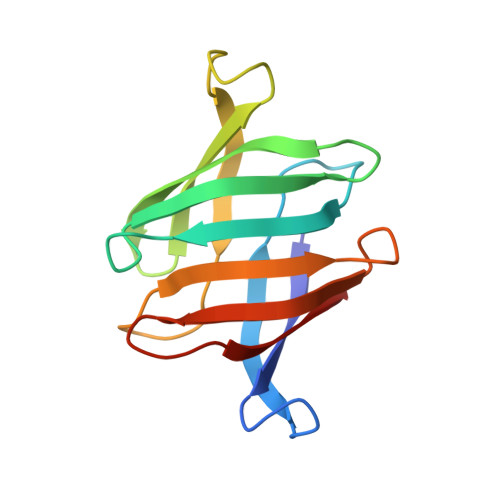
Structural basis of the anti-HIV activity of the cyanobacterial Oscillatoria Agardhii agglutinin.

Koharudin, L.M.Gronenborn, A.M.

(2011) Structure 19: 1170-1181

  • DOI: https://doi.org/10.1016/j.str.2011.05.010
  • Primary Citation Related Structures: 
    3S5V, 3S5X, 3S60

  • PubMed Abstract: 

    The cyanobacterial Oscillatory Agardhii agglutinin (OAA) is a recently discovered HIV-inactivating lectin that interacts with high-mannose sugars. Nuclear magnetic resonance (NMR) binding studies between OAA and α3,α6-mannopentaose (Manα(1-3)[Manα(1-3)[Manα(1-6)]Manα(1-6)]Man), the branched core unit of Man-9, revealed two binding sites at opposite ends of the protein, exhibiting essentially identical affinities. Atomic details of the specific protein-sugar contacts in the recognition loops of OAA were delineated in the high-resolution crystal structures of free and glycan-complexed protein. No major changes in the overall protein structure are induced by carbohydrate binding, with essentially identical apo- and sugar-bound conformations in binding site 1. A single peptide bond flip at W77-G78 is seen in binding site 2. Our combined NMR and crystallographic results provide structural insights into the mechanism by which OAA specifically recognizes the branched Man-9 core, distinctly different from the recognition of the D1 and D3 arms at the nonreducing end of high-mannose carbohydrates by other antiviral lectins.


  • Organizational Affiliation
    • Department of Structural Biology, School of Medicine, University of Pittsburgh, Biomedical Science Tower 3, Pittsburgh, PA 15260, USA.

Macromolecule Content 

  • Total Structure Weight: 14.06 kDa 
  • Atom Count: 1,066 
  • Modeled Residue Count: 132 
  • Deposited Residue Count: 133 
  • Unique protein chains: 1

Macromolecules

Find similar proteins by:|  3D Structure
Entity ID: 1
MoleculeChains  Sequence LengthOrganismDetailsImage
Lectin133Planktothrix agardhiiMutation(s): 0 
Gene Names: OAA
UniProt
Find proteins for P84330 (Planktothrix agardhii)
Explore P84330 
Go to UniProtKB:  P84330
Entity Groups
Sequence Clusters30% Identity50% Identity70% Identity90% Identity95% Identity100% Identity
UniProt GroupP84330
Sequence Annotations
Expand
Reference Sequence

Experimental Data & Validation

Experimental Data

  • Method: X-RAY DIFFRACTION
  • Resolution: 1.60 Å
  • R-Value Free:  0.228 (Depositor), 0.239 (DCC) 
  • R-Value Work:  0.182 (Depositor), 0.192 (DCC) 
  • R-Value Observed: 0.187 (Depositor) 
Space Group: P 21 21 21
Unit Cell:
Length ( Å )Angle ( ˚ )
a = 39.467α = 90
b = 40.658β = 90
c = 70.374γ = 90
Software Package:
Software NamePurpose
StructureStudiodata collection
PHASERphasing
REFMACrefinement
d*TREKdata reduction
d*TREKdata scaling

Structure Validation

View Full Validation Report


Entry History 

Deposition Data

Revision History  (Full details and data files)

  • Version 1.0: 2011-06-15
    Type: Initial release
  • Version 1.1: 2011-07-13
    Changes: Version format compliance
  • Version 1.2: 2012-04-25
    Changes: Database references
  • Version 1.3: 2023-09-13
    Changes: Data collection, Database references, Refinement description