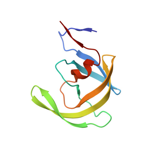
Critical differences in HIV-1 and HIV-2 protease specificity for clinical inhibitors.
Tie, Y., Wang, Y.F., Boross, P.I., Chiu, T.Y., Ghosh, A.K., Tozser, J., Louis, J.M., Harrison, R.W., Weber, I.T.(2012) Protein Sci 21: 339-350
- PubMed: 22238126
- DOI: https://doi.org/10.1002/pro.2019
- Primary Citation of Related Structures:
3S43, 3S45, 3S53, 3S54, 3S56 - PubMed Abstract:
Clinical inhibitor amprenavir (APV) is less effective on HIV-2 protease (PR₂) than on HIV-1 protease (PR₁). We solved the crystal structure of PR₂ with APV at 1.5 Å resolution to identify structural changes associated with the lowered inhibition. Furthermore, we analyzed the PR₁ mutant (PR(1M) ) with substitutions V32I, I47V, and V82I that mimic the inhibitor binding site of PR₂. PR(1M) more closely resembled PR₂ than PR₁ in catalytic efficiency on four substrate peptides and inhibition by APV, whereas few differences were seen for two other substrates and inhibition by saquinavir (SQV) and darunavir (DRV). High resolution crystal structures of PR(1M) with APV, DRV, and SQV were compared with available PR₁ and PR₂ complexes. Val/Ile32 and Ile/Val47 showed compensating interactions with SQV in PR(1M) and PR₁, however, Ile82 interacted with a second SQV bound in an extension of the active site cavity of PR(1M). Residues 32 and 82 maintained similar interactions with DRV and APV in all the enzymes, whereas Val47 and Ile47 had opposing effects in the two subunits. Significantly diminished interactions were seen for the aniline of APV bound in PR₁ (M) and PR₂ relative to the strong hydrogen bonds observed in PR₁, consistent with 15- and 19-fold weaker inhibition, respectively. Overall, PR(1M) partially replicates the specificity of PR₂ and gives insight into drug resistant mutations at residues 32, 47, and 82. Moreover, this analysis provides a structural explanation for the weaker antiviral effects of APV on HIV-2.
Organizational Affiliation:
Department of Biology, Georgia State University, Atlanta, Georgia 30303, USA.


