3RX9 | pdb_00003rx9

3D structure of SciN from an Escherichia coli Patotype


Experimental Data Snapshot

  • Method: X-RAY DIFFRACTION
  • Resolution: 1.35 Å
  • R-Value Free: 
    0.160 (Depositor), 0.161 (DCC) 
  • R-Value Work: 
    0.137 (Depositor) 
  • R-Value Observed: 
    0.138 (Depositor) 

Starting Model: experimental
View more details

wwPDB Validation 3D Report Full Report

Validation slider image for 3RX9

This is version 1.2 of the entry. See complete history

Literature

Towards a Structural Comprehension of Bacterial Type VI Secretion Systems: Characterization of the TssJ-TssM Complex of an Escherichia coli Pathovar.

Felisberto-Rodrigues, C.Durand, E.Aschtgen, M.S.Blangy, S.Ortiz-Lombardia, M.Douzi, B.Cambillau, C.Cascales, E.

(2011) PLoS Pathog 7: e1002386-e1002386

  • DOI: https://doi.org/10.1371/journal.ppat.1002386
  • Primary Citation Related Structures: 
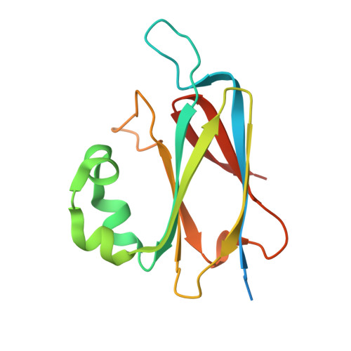
    3RX9

  • PubMed Abstract: 

    Type VI secretion systems (T6SS) are trans-envelope machines dedicated to the secretion of virulence factors into eukaryotic or prokaryotic cells, therefore required for pathogenesis and/or for competition towards neighboring bacteria. The T6SS apparatus resembles the injection device of bacteriophage T4, and is anchored to the cell envelope through a membrane complex. This membrane complex is composed of the TssL, TssM and TagL inner membrane anchored proteins and of the TssJ outer membrane lipoprotein. Here, we report the crystal structure of the enteroaggregative Escherichia coli Sci1 TssJ lipoprotein, a two four-stranded β-sheets protein that exhibits a transthyretin fold with an additional α-helical domain and a protruding loop. We showed that TssJ contacts TssM through this loop since a loop depleted mutant failed to interact with TssM in vitro or in vivo. Biophysical analysis of TssM and TssJ-TssM interaction suggest a structural model of the membrane-anchored outer shell of T6SS. Collectively, our results provide an improved understanding of T6SS assembly and encourage structure-aided drug design of novel antimicrobials targeting T6SS.


  • Organizational Affiliation
    • Aix-Marseille Université, Architecture et Fonction des Macromolécules Biologiques, Campus de Luminy, Marseille, France.

Macromolecule Content 

  • Total Structure Weight: 17.07 kDa 
  • Atom Count: 1,366 
  • Modeled Residue Count: 130 
  • Deposited Residue Count: 155 
  • Unique protein chains: 1

Macromolecules

Find similar proteins by:|  3D Structure
Entity ID: 1
MoleculeChains  Sequence LengthOrganismDetailsImage
Putative uncharacterized protein155Escherichia coli 55989Mutation(s): 0 
Gene Names: EC55989_3320NC_011748.1
UniProt
Find proteins for B7LFS8 (Escherichia coli (strain 55989 / EAEC))
Explore B7LFS8 
Go to UniProtKB:  B7LFS8
Entity Groups
Sequence Clusters30% Identity50% Identity70% Identity90% Identity95% Identity100% Identity
UniProt GroupB7LFS8
Sequence Annotations
Expand
Reference Sequence

Small Molecules

Ligands 1 Unique
IDChains Name / Formula / InChI Key2D Diagram3D Interactions
SO4

Query on SO4


Download:Ideal Coordinates CCD File
B [auth A]SULFATE ION
O4 S
QAOWNCQODCNURD-UHFFFAOYSA-L

Experimental Data & Validation

Experimental Data

  • Method: X-RAY DIFFRACTION
  • Resolution: 1.35 Å
  • R-Value Free:  0.160 (Depositor), 0.161 (DCC) 
  • R-Value Work:  0.137 (Depositor) 
  • R-Value Observed: 0.138 (Depositor) 
Space Group: P 31 2 1
Unit Cell:
Length ( Å )Angle ( ˚ )
a = 78.07α = 90
b = 78.07β = 90
c = 46.98γ = 120
Software Package:
Software NamePurpose
ADSCdata collection
MOLREPphasing
REFMACrefinement
XDSdata reduction
XSCALEdata scaling

Structure Validation

View Full Validation Report


Entry History 

Deposition Data

Revision History  (Full details and data files)

  • Version 1.0: 2011-12-07
    Type: Initial release
  • Version 1.1: 2024-02-28
    Changes: Data collection, Database references, Derived calculations
  • Version 1.2: 2024-04-03
    Changes: Refinement description