


Crystal structure of complex of 4PAL (4-Pyridoxolactone) and PLDH (tetrameric pyridoxal 4-dehydrogenase) from Mesorhizobium loti
Yagi, T., Chu, H.N.To be published.
Experimental Data Snapshot
Starting Model: experimental
View more details
Entity ID: 1 | |||||
|---|---|---|---|---|---|
| Molecule | Chains | Sequence Length | Organism | Details | Image |
| Pyridoxal 4-dehydrogenase | 247 | Mesorhizobium loti | Mutation(s): 0 Gene Names: mlr6807, pldh-t EC: 1.1.1.107 | 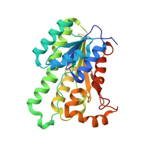 | |
UniProt | |||||
Entity Groups | |||||
| Sequence Clusters | 30% Identity50% Identity70% Identity90% Identity95% Identity100% Identity | ||||
| UniProt Group | Q988B7 | ||||
Sequence AnnotationsExpand | |||||
Reference Sequence | |||||
| Ligands 3 Unique | |||||
|---|---|---|---|---|---|
| ID | Chains | Name / Formula / InChI Key | 2D Diagram | 3D Interactions | |
| NAD Download:Ideal Coordinates CCD File | E [auth A], J [auth B], L [auth C], O [auth D] | NICOTINAMIDE-ADENINE-DINUCLEOTIDE C21 H27 N7 O14 P2 BAWFJGJZGIEFAR-NNYOXOHSSA-N |  | ||
| 4PL Download:Ideal Coordinates CCD File | F [auth A], K [auth B], M [auth C], P [auth D] | 7-hydroxy-6-methylfuro[3,4-c]pyridin-1(3H)-one C8 H7 N O3 HHPDVQLBYQFYFA-UHFFFAOYSA-N |  | ||
| GOL Download:Ideal Coordinates CCD File | G [auth A], H [auth A], I [auth A], N [auth C], Q [auth D] | GLYCEROL C3 H8 O3 PEDCQBHIVMGVHV-UHFFFAOYSA-N |  | ||
| Length ( Å ) | Angle ( ˚ ) |
|---|---|
| a = 85.537 | α = 90 |
| b = 50.211 | β = 90.47 |
| c = 94.266 | γ = 90 |
| Software Name | Purpose |
|---|---|
| HKL-2000 | data collection |
| PHASES | phasing |
| PHENIX | refinement |
| HKL-2000 | data reduction |
| HKL-2000 | data scaling |


