3RV5

Crystal structure of human cardiac troponin C regulatory domain in complex with cadmium and deoxycholic acid


Experimental Data Snapshot

  • Method: X-RAY DIFFRACTION
  • Resolution: 2.20 Å
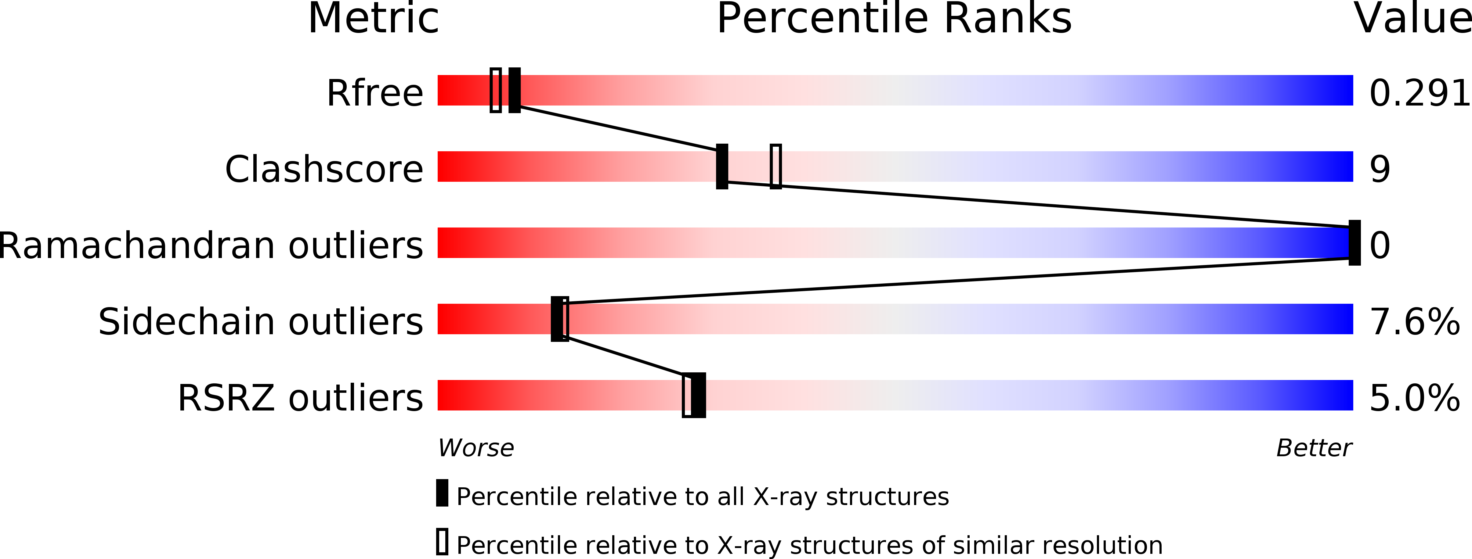
  • R-Value Free: 0.281 
  • R-Value Work: 0.225 
  • R-Value Observed: 0.228 

wwPDB Validation   3D Report Full Report


Ligand Structure Quality Assessment 


This is version 1.3 of the entry. See complete history


Literature

Crystal structure of cardiac troponin C regulatory domain in complex with cadmium and deoxycholic Acid reveals novel conformation.

Li, A.Y.Lee, J.Borek, D.Otwinowski, Z.Tibbits, G.F.Paetzel, M.

(2011) J Mol Biol 413: 699-711

  • DOI: https://doi.org/10.1016/j.jmb.2011.08.049
  • Primary Citation of Related Structures:  
    3RV5

  • PubMed Abstract: 

    The amino-terminal regulatory domain of cardiac troponin C (cNTnC) plays an important role as the calcium sensor for the troponin complex. Calcium binding to cNTnC results in conformational changes that trigger a cascade of events that lead to cardiac muscle contraction. The cardiac N-terminal domain of TnC consists of two EF-hand calcium binding motifs, one of which is dysfunctional in binding calcium. Nevertheless, the defunct EF-hand still maintains a role in cNTnC function. For its structural analysis by X-ray crystallography, human cNTnC with the wild-type primary sequence was crystallized under a novel crystallization condition. The crystal structure was solved by the single-wavelength anomalous dispersion method and refined to 2.2 Å resolution. The structure displays several novel features. Firstly, both EF-hand motifs coordinate cadmium ions derived from the crystallization milieu. Secondly, the ion coordination in the defunct EF-hand motif accompanies unusual changes in the protein conformation. Thirdly, deoxycholic acid, also derived from the crystallization milieu, is bound in the central hydrophobic cavity. This is reminiscent of the interactions observed for cardiac calcium sensitizer drugs that bind to the same core region and maintain the "open" conformational state of calcium-bound cNTnC. The cadmium ion coordination in the defunct EF-hand indicates that this vestigial calcium binding site retains the structural and functional elements that allow it to coordinate a cadmium ion. However, it is a result of, or concomitant with, large and unusual structural changes in cNTnC.


  • Organizational Affiliation

    Department of Molecular Biology and Biochemistry, Simon Fraser University, South Science Building, 8888 University Drive, Burnaby, British Columbia, Canada, V5A 1S6.


Macromolecules
Find similar proteins by:  (by identity cutoff)  |  3D Structure
Entity ID: 1
MoleculeChains Sequence LengthOrganismDetailsImage
Troponin C, slow skeletal and cardiac muscles
A, B, C, D
89Homo sapiensMutation(s): 0 
Gene Names: TNNCTNNC1TNNC1 (Amino acids 1-89)
UniProt & NIH Common Fund Data Resources
Find proteins for P63316 (Homo sapiens)
Explore P63316 
Go to UniProtKB:  P63316
PHAROS:  P63316
GTEx:  ENSG00000114854 
Entity Groups  
Sequence Clusters30% Identity50% Identity70% Identity90% Identity95% Identity100% Identity
UniProt GroupP63316
Sequence Annotations
Expand
  • Reference Sequence
Small Molecules
Ligands 3 Unique
IDChains Name / Formula / InChI Key2D Diagram3D Interactions
DXC
Query on DXC

Download Ideal Coordinates CCD File 
BA [auth C],
GA [auth D],
J [auth A],
K [auth A],
L [auth B]
(3ALPHA,5BETA,12ALPHA)-3,12-DIHYDROXYCHOLAN-24-OIC ACID
C24 H40 O4
KXGVEGMKQFWNSR-LLQZFEROSA-N
CD
Query on CD

Download Ideal Coordinates CCD File 
CA [auth D]
DA [auth D]
E [auth A]
EA [auth D]
F [auth A]
CA [auth D],
DA [auth D],
E [auth A],
EA [auth D],
F [auth A],
FA [auth D],
G [auth A],
H [auth A],
I [auth A],
O [auth B],
P [auth B],
Q [auth B],
R [auth B],
S [auth B],
T [auth B],
U [auth B],
V [auth C],
W [auth C],
X [auth C],
Y [auth C],
Z [auth C]
CADMIUM ION
Cd
WLZRMCYVCSSEQC-UHFFFAOYSA-N
CA
Query on CA

Download Ideal Coordinates CCD File 
AA [auth C],
M [auth B],
N [auth B]
CALCIUM ION
Ca
BHPQYMZQTOCNFJ-UHFFFAOYSA-N
Modified Residues  1 Unique
IDChains TypeFormula2D DiagramParent
MSE
Query on MSE
A, B, C, D
L-PEPTIDE LINKINGC5 H11 N O2 SeMET
Experimental Data & Validation

Experimental Data

  • Method: X-RAY DIFFRACTION
  • Resolution: 2.20 Å
  • R-Value Free: 0.281 
  • R-Value Work: 0.225 
  • R-Value Observed: 0.228 
  • Space Group: P 21 21 2
Unit Cell:
Length ( Å )Angle ( ˚ )
a = 51.844α = 90
b = 81.783β = 90
c = 100.469γ = 90
Software Package:
Software NamePurpose
HKL-2000data collection
SHELXSphasing
REFMACrefinement
HKL-2000data reduction
HKL-2000data scaling

Structure Validation

View Full Validation Report


Ligand Structure Quality Assessment 


Entry History 

Deposition Data

Revision History  (Full details and data files)

  • Version 1.0: 2011-08-31
    Type: Initial release
  • Version 1.1: 2011-10-26
    Changes: Database references
  • Version 1.2: 2011-11-16
    Changes: Database references
  • Version 1.3: 2024-10-16
    Changes: Data collection, Database references, Derived calculations, Structure summary