3RTV | pdb_00003rtv

Crystal structure of the large fragment of DNA polymerase I from Thermus Aquaticus in a closed ternary complex with natural primer/template DNA


Experimental Data Snapshot

  • Method: X-RAY DIFFRACTION
  • Resolution: 1.90 Å
  • R-Value Free: 
    0.202 (Depositor), 0.199 (DCC) 
  • R-Value Work: 
    0.162 (Depositor), 0.159 (DCC) 
  • R-Value Observed: 
    0.164 (Depositor) 

Starting Model: experimental
View more details

wwPDB Validation 3D Report Full Report

Validation slider image for 3RTV

Ligand Structure Quality Assessment 


This is version 1.3 of the entry. See complete history

Literature

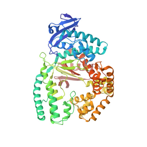
KlenTaq polymerase replicates unnatural base pairs by inducing a Watson-Crick geometry.

Betz, K.Malyshev, D.A.Lavergne, T.Welte, W.Diederichs, K.Dwyer, T.J.Ordoukhanian, P.Romesberg, F.E.Marx, A.

(2012) Nat Chem Biol 8: 612-614

  • DOI: https://doi.org/10.1038/nchembio.966
  • Primary Citation Related Structures: 
    3RTV, 3SV3, 3SV4, 3SYZ, 3SZ2

  • PubMed Abstract: 

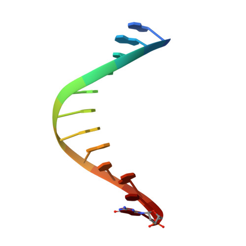
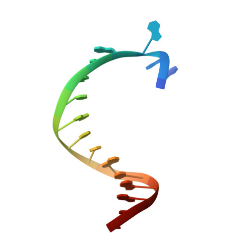
    Many candidate unnatural DNA base pairs have been developed, but some of the best-replicated pairs adopt intercalated structures in free DNA that are difficult to reconcile with known mechanisms of polymerase recognition. Here we present crystal structures of KlenTaq DNA polymerase at different stages of replication for one such pair, dNaM-d5SICS, and show that efficient replication results from the polymerase itself, inducing the required natural-like structure.


  • Organizational Affiliation
    • Department of Chemistry, Universität Konstanz, Konstanz, Germany.

Macromolecule Content 

  • Total Structure Weight: 70.79 kDa 
  • Atom Count: 5,354 
  • Modeled Residue Count: 568 
  • Deposited Residue Count: 568 
  • Unique protein chains: 1
  • Unique nucleic acid chains: 2

Macromolecules


Find similar proteins by:|  3D Structure
Entity ID: 1
MoleculeChains  Sequence LengthOrganismDetailsImage
DNA polymerase I, thermostable540Thermus aquaticusMutation(s): 0 
Gene Names: pol Ipol1polA
EC: 2.7.7.7
UniProt
Find proteins for P19821 (Thermus aquaticus)
Explore P19821 
Go to UniProtKB:  P19821
Entity Groups
Sequence Clusters30% Identity50% Identity70% Identity90% Identity95% Identity100% Identity
UniProt GroupP19821
Sequence Annotations
Expand
Reference Sequence
Find similar nucleic acids by:  Sequence
Entity ID: 2
MoleculeChains LengthOrganismImage
(5'-D(*GP*AP*CP*CP*AP*CP*GP*GP*CP*GP*CP*(DDG))-3')12N/A
Sequence Annotations
Expand
Reference Sequence
Find similar nucleic acids by:  Sequence
Entity ID: 3
MoleculeChains LengthOrganismImage
(5'-D(*AP*AP*AP*GP*CP*GP*CP*GP*CP*CP*GP*TP*GP*GP*TP*C)-3')16N/A
Sequence Annotations
Expand
Reference Sequence

Small Molecules

Ligands 4 Unique
IDChains Name / Formula / InChI Key2D Diagram3D Interactions
DCP

Query on DCP


Download:Ideal Coordinates CCD File
D [auth A]2'-DEOXYCYTIDINE-5'-TRIPHOSPHATE
C9 H16 N3 O13 P3
RGWHQCVHVJXOKC-SHYZEUOFSA-N
GOL

Query on GOL


Download:Ideal Coordinates CCD File
G [auth A]
H [auth A]
I [auth A]
J [auth A]
N [auth A]
G [auth A],
H [auth A],
I [auth A],
J [auth A],
N [auth A],
P [auth C]
GLYCEROL
C3 H8 O3
PEDCQBHIVMGVHV-UHFFFAOYSA-N
ACT

Query on ACT


Download:Ideal Coordinates CCD File
K [auth A],
L [auth A],
M [auth A]
ACETATE ION
C2 H3 O2
QTBSBXVTEAMEQO-UHFFFAOYSA-M
MG

Query on MG


Download:Ideal Coordinates CCD File
E [auth A],
F [auth A],
O [auth B]
MAGNESIUM ION
Mg
JLVVSXFLKOJNIY-UHFFFAOYSA-N

Experimental Data & Validation

Experimental Data

  • Method: X-RAY DIFFRACTION
  • Resolution: 1.90 Å
  • R-Value Free:  0.202 (Depositor), 0.199 (DCC) 
  • R-Value Work:  0.162 (Depositor), 0.159 (DCC) 
  • R-Value Observed: 0.164 (Depositor) 
Space Group: P 31 2 1
Unit Cell:
Length ( Å )Angle ( ˚ )
a = 108.296α = 90
b = 108.296β = 90
c = 90.381γ = 120
Software Package:
Software NamePurpose
PHENIXmodel building
PHENIXrefinement
XDSdata reduction
XDSdata scaling
PHENIXphasing

Structure Validation

View Full Validation Report


Ligand Structure Quality Assessment 


Entry History 

Deposition Data

Revision History  (Full details and data files)

  • Version 1.0: 2012-05-30
    Type: Initial release
  • Version 1.1: 2012-07-04
    Changes: Database references
  • Version 1.2: 2012-09-12
    Changes: Database references
  • Version 1.3: 2023-09-13
    Changes: Data collection, Database references, Derived calculations, Refinement description