3RPS

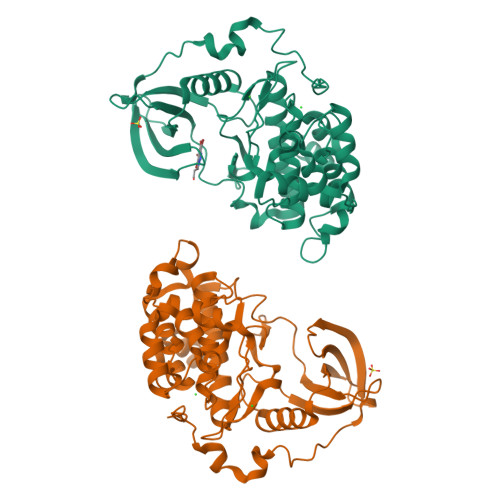
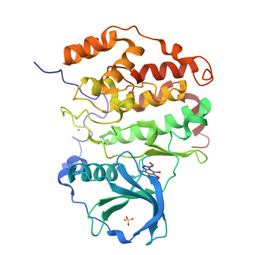
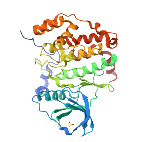
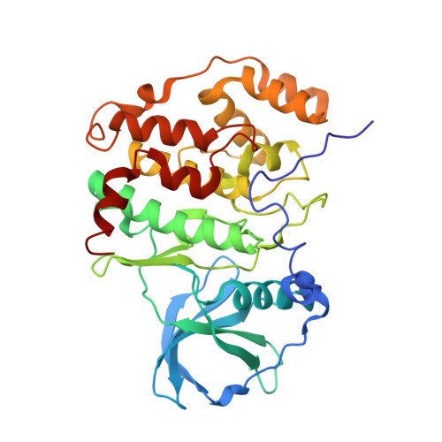
Structure of human CK2alpha in complex with the ATP-competitive inhibitor 3-(4,5,6,7-tetrabromo-1H-benzotriazol-1-yl)propan-1-ol


Experimental Data Snapshot

  • Method: X-RAY DIFFRACTION
  • Resolution: 2.30 Å
  • R-Value Free: 0.228 
  • R-Value Work: 0.170 
  • R-Value Observed: 0.172 

wwPDB Validation   3D Report Full Report


Ligand Structure Quality Assessment 

Created with Raphaël 2.3.0Worse 01 BetterLigand structure goodness of fit to experimental dataBest fitted 4B0Click on this verticalbar to view details

This is version 1.3 of the entry. See complete history


Literature

Enzymatic activity with an incomplete catalytic spine - insights from a comparative structural analysis of human CK2alpha and its paralogous isoform CK2alpha'

Bischoff, N.Raaf, J.Olsen, B.Bretner, M.Issinger, O.G.Niefind, K.

(2011) Mol Cell Biochem 356: 57-65

  • DOI: https://doi.org/10.1007/s11010-011-0948-5
  • Primary Citation of Related Structures:  
    3RPS

  • PubMed Abstract: 

    Eukaryotic protein kinases are fundamental factors for cellular regulation and therefore subject of strict control mechanisms. For full activity a kinase molecule must be penetrated by two stacks of hydrophobic residues, the regulatory and the catalytic spine that are normally well conserved among active protein kinases. We apply this novel spine concept here on CK2α, the catalytic subunit of protein kinase CK2. Homo sapiens disposes of two paralog isoforms of CK2α (hsCK2α and hsCK2α'). We describe two new structures of hsCK2α constructs one of which in complex with the ATP-analog adenylyl imidodiphosphate and the other with the ATP-competitive inhibitor 3-(4,5,6,7-tetrabromo-1H-benzotriazol-1-yl)propan-1-ol. The former is the first hsCK2α structure with a well defined cosubstrate/magnesium complex and the second with an open β4/β5-loop. Comparisons of these structures with existing CK2α/CK2α' and cAMP-dependent protein kinase (PKA) structures reveal: in hsCK2α' an open conformation of the interdomain hinge/helix αD region that is critical for ATP-binding is found corresponding to an incomplete catalytic spine. In contrast hsCK2α often adopts the canonical, PKA-like version of the catalytic spine which correlates with a closed conformation of the hinge region. HsCK2α can switch to the incomplete, non-canonical, hsCK2α'-like state of the catalytic spine, but this transition apparently depends on binding of either ATP or of the regulatory subunit CK2β. Thus, ATP looks like an activator of hsCK2α rather than a pure cosubstrate.


  • Organizational Affiliation

    Universität zu Köln, Institut für Biochemie, Zülpicher Straße 47, 50674 Köln, Germany.


Macromolecules
Find similar proteins by:  (by identity cutoff)  |  3D Structure
Entity ID: 1
MoleculeChains Sequence LengthOrganismDetailsImage
Casein kinase II subunit alpha
A, B
335Homo sapiensMutation(s): 0 
Gene Names: CSNK2A1CK2A1
EC: 2.7.11.1
UniProt & NIH Common Fund Data Resources
Find proteins for P68400 (Homo sapiens)
Explore P68400 
Go to UniProtKB:  P68400
PHAROS:  P68400
GTEx:  ENSG00000101266 
Entity Groups  
Sequence Clusters30% Identity50% Identity70% Identity90% Identity95% Identity100% Identity
UniProt GroupP68400
Sequence Annotations
Expand
  • Reference Sequence
Small Molecules
Ligands 3 Unique
IDChains Name / Formula / InChI Key2D Diagram3D Interactions
4B0
Query on 4B0

Download Ideal Coordinates CCD File 
C [auth A]3-(4,5,6,7-tetrabromo-1H-benzotriazol-1-yl)propan-1-ol
C9 H7 Br4 N3 O
VXLNELSTYPRMEW-UHFFFAOYSA-N
SO4
Query on SO4

Download Ideal Coordinates CCD File 
D [auth A],
G [auth B]
SULFATE ION
O4 S
QAOWNCQODCNURD-UHFFFAOYSA-L
CL
Query on CL

Download Ideal Coordinates CCD File 
E [auth A],
F [auth A],
H [auth B],
I [auth B]
CHLORIDE ION
Cl
VEXZGXHMUGYJMC-UHFFFAOYSA-M
Binding Affinity Annotations 
IDSourceBinding Affinity
4B0 BindingDB:  3RPS IC50: 320 (nM) from 1 assay(s)
Experimental Data & Validation

Experimental Data

  • Method: X-RAY DIFFRACTION
  • Resolution: 2.30 Å
  • R-Value Free: 0.228 
  • R-Value Work: 0.170 
  • R-Value Observed: 0.172 
  • Space Group: P 43
Unit Cell:
Length ( Å )Angle ( ˚ )
a = 72.01α = 90
b = 72.01β = 90
c = 133.12γ = 90
Software Package:
Software NamePurpose
PHASERphasing
REFMACrefinement
XDSdata reduction
XSCALEdata scaling

Structure Validation

View Full Validation Report


Ligand Structure Quality Assessment 

Created with Raphaël 2.3.0Worse 01 BetterLigand structure goodness of fit to experimental dataBest fitted 4B0Click on this verticalbar to view details

Entry History 

Deposition Data

Revision History  (Full details and data files)

  • Version 1.0: 2011-06-08
    Type: Initial release
  • Version 1.1: 2011-07-13
    Changes: Version format compliance
  • Version 1.2: 2013-08-21
    Changes: Database references
  • Version 1.3: 2024-02-28
    Changes: Data collection, Database references, Derived calculations