3ROH | pdb_00003roh

Crystal Structure of Leukotoxin (LukE) from Staphylococcus aureus subsp. aureus COL.


Experimental Data Snapshot

  • Method: X-RAY DIFFRACTION
  • Resolution: 3.20 Å
  • R-Value Free: 
    0.224 (Depositor), 0.227 (DCC) 
  • R-Value Work: 
    0.178 (Depositor), 0.182 (DCC) 
  • R-Value Observed: 
    0.181 (Depositor) 

Starting Model: experimental
View more details

wwPDB Validation 3D Report Full Report

Validation slider image for 3ROH

This is version 1.5 of the entry. See complete history

Literature

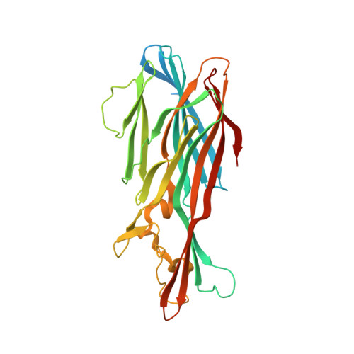
Crystal structures of the components of the Staphylococcus aureus leukotoxin ED.

Nocadello, S.Minasov, G.Shuvalova, L.Dubrovska, I.Sabini, E.Bagnoli, F.Grandi, G.Anderson, W.F.

(2016) Acta Crystallogr D Biol Crystallogr 72: 113-120

  • DOI: https://doi.org/10.1107/S2059798315023207
  • Primary Citation Related Structures: 
    3ROH, 4Q7G

  • PubMed Abstract: 

    Staphylococcal leukotoxins are a family of β-barrel, bicomponent, pore-forming toxins with membrane-damaging functions. These bacterial exotoxins share sequence and structural homology and target several host-cell types. Leukotoxin ED (LukED) is one of these bicomponent pore-forming toxins that Staphylococcus aureus produces in order to suppress the ability of the host to contain the infection. The recent delineation of the important role that LukED plays in S. aureus pathogenesis and the identification of its protein receptors, combined with its presence in S. aureus methicillin-resistant epidemic strains, establish this leukocidin as a possible target for the development of novel therapeutics. Here, the crystal structures of the water-soluble LukE and LukD components of LukED have been determined. The two structures illustrate the tertiary-structural variability with respect to the other leukotoxins while retaining the conservation of the residues involved in the interaction of the protomers in the bipartite leukotoxin in the pore complex.


  • Organizational Affiliation
    • Center for Structural Genomics of Infectious Diseases, Department of Biochemistry and Molecular Genetics, Northwestern University Feinberg School of Medicine, Chicago, IL 60611, USA.

Macromolecule Content 

  • Total Structure Weight: 37.26 kDa 
  • Atom Count: 2,286 
  • Modeled Residue Count: 282 
  • Deposited Residue Count: 329 
  • Unique protein chains: 1

Macromolecules

Find similar proteins by:|  3D Structure
Entity ID: 1
MoleculeChains  Sequence LengthOrganismDetailsImage
Leucotoxin LukEv329Staphylococcus aureus subsp. aureus NCTC 8325Mutation(s): 0 
Gene Names: HMPREF0773_11197lukElukEvSAOUHSC_01955
UniProt
Find proteins for Q2FXB0 (Staphylococcus aureus (strain NCTC 8325 / PS 47))
Explore Q2FXB0 
Go to UniProtKB:  Q2FXB0
Entity Groups
Sequence Clusters30% Identity50% Identity70% Identity90% Identity95% Identity100% Identity
UniProt GroupQ2FXB0
Sequence Annotations
Expand
Reference Sequence

Experimental Data & Validation

Experimental Data

  • Method: X-RAY DIFFRACTION
  • Resolution: 3.20 Å
  • R-Value Free:  0.224 (Depositor), 0.227 (DCC) 
  • R-Value Work:  0.178 (Depositor), 0.182 (DCC) 
  • R-Value Observed: 0.181 (Depositor) 
Space Group: I 4
Unit Cell:
Length ( Å )Angle ( ˚ )
a = 133.904α = 90
b = 133.904β = 90
c = 64.473γ = 90
Software Package:
Software NamePurpose
Blu-Icedata collection
PHASERphasing
REFMACrefinement
HKL-2000data reduction
HKL-2000data scaling

Structure Validation

View Full Validation Report


Entry History 

Revision History  (Full details and data files)

  • Version 1.0: 2011-05-04
    Type: Initial release
  • Version 1.1: 2011-07-13
    Changes: Version format compliance
  • Version 1.2: 2016-03-02
    Changes: Database references
  • Version 1.3: 2016-03-16
    Changes: Source and taxonomy
  • Version 1.4: 2017-11-08
    Changes: Refinement description
  • Version 1.5: 2023-09-13
    Changes: Data collection, Database references, Derived calculations, Refinement description